
- Title
-
Kit signaling is involved in melanocyte stem cell fate decisions in zebrafish embryos
- Authors
- O'Reilly-Pol, T., and Johnson, S.L.
- Source
- Full text @ Development

Melanocyte regeneration is more sensitive to deficits in Kit signaling than ontogeny. (A) Schema of the experiments. Zebrafish were reared until 3 dpf and scored for ontogenetic melanocytes (open arrowhead) or treated with 4-HA from 1-3 dpf and scored for regeneration melanocytes at 6 dpf (filled arrowhead). (B) Comparison of kita mutant genotypes to WT. Three to ten embryos were compared with WT controls for ontogenetic melanocytes (white bars) and regeneration melanocytes (black bars). Experiments with kitats were performed at 25°C, and all others were performed at 28.5°C. Error bars indicate s.d. *P<0.05, **P<0.001. PHENOTYPE:
|
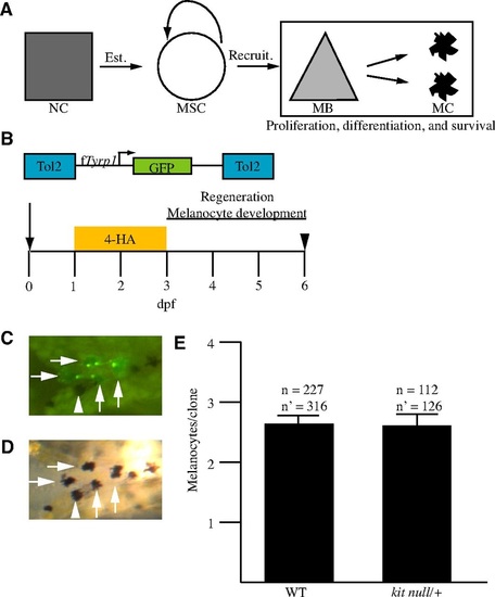
MSC daughter cell proliferation, differentiation and survival are unaffected in kitanull/+ embryos. (A) A general model of melanocyte regeneration. A neural crest progenitor (square, NC) establishes a melanocyte stem cell (circle, MSC), which can be recruited to produce melanoblasts (triangle, MB), which will proliferate and differentiate into melanocytes (MC). Box indicates process tested. (B) Schema of the experiment. Zebrafish embryos were injected at the one- to two-cell stage (arrow) with transposase and a transposon containing fTyrp1>GFP, treated with 4-HA from 1-3 dpf to ablate ontogenetic melanocytes, and scored for regeneration melanocytes at day 6 (arrowhead). (C,D) Example of a clone in WT. (C) An epifluorescent image of an embryo containing a clone. (D) A brightfield image of the embryo shown in C. Melanocytes containing GFP are marked with an arrow. Some melanocytes lack GFP, and one example is marked with an arrowhead. (E) The mean number of melanocytes per clone is the same for both genotypes in regeneration. n, the number of embryos containing clones; n′ the number of clones after adjustment for polyclonal events. Error bars are s.e.m. |

Any reduction in kit signaling affects melanocyte regeneration. (A) Schema of the experiments. Fish were reared until 3 dpf and scored for ontogenetic melanocytes (open arrowhead) or treated with 4-HA from 1-3 dpf and scored for regeneration melanocytes at 6 dpf (filled arrowhead). (B) Comparison of kita mutant genotypes with WT. Three to ten embryos were compared with WT controls for ontogenetic melanocytes (empty bars) and regeneration melanocytes (filled bars). Alleles kitab5 and kitaj1e99 are referred to as kitanull and kitats in the main text and figures. Allele kitaj1e60 affects ontogenetic melanocyte migration without affecting ontogenetic melanocyte survival (Rawls and Johnson, 2003). Allele kitaj1e78 affects ontogenetic melanocyte survival without affecting ontogenetic melanocyte migration (Rawls and Johnson, 2003). Experiments with kitaj1e99 were performed at 25°C, and all others were performed at 28.5°C. Error bars indicate s.d. *P<0.05, **P<0.005, ***P<0.001. PHENOTYPE:
|