
- Title
-
Revealing details: whole mount microRNA in situ hybridization protocol for zebrafish embryos and adult tissues
- Authors
- Lagendijk, A.K., Moulton, J.D., and Bakkers, J.
- Source
- Full text @ Biol. Open

Whole mount miRNA in situ hybridization by CF-labeled MO probes. (A–B) In situ hybridization using a CF-labeled miR-124 MO probe on PFA fixed 4 dpf embryo detects expression in developing brain and eyes (A, dorsal view; B, lateral view). (C–D) In situ hybridization using a CF-labeled miR-206 MO probe on PFA fixed 4 dpf embryo detects expression of miR-206 in skeletal muscle cells (C, dorsal view; D, lateral view). |

Whole mount miRNA in situ hybridization on embryos. Top rows of top and bottom panel: miRNA in situ hybridization on embryos fixed in PFA only. Bottom rows of top and bottom panel: miRNA expression detected in embryos fixed in PFA+EDC. All embryos where processed simultaneously and stained for approximately 24 h. All arrowheads indicate miRNA expression detectable with high resolution in PFA+EDC fixed samples versus fixation in PFA only. Asterisks indicate non-specific background signals in PFA fixed embryos (A,E,G,K,Q). (A–D) MiR-206 expression in skeletal muscle cells was detected by a CF-labeled MO probe at 2 dpf (A,B) and 3 dpf (C,D). Arrowheads (A,B) point to skeletal muscle cells located on the yolk that were detectable in PFA+EDC fixed embryos (B) and not in PFA fixed embryos (A). (E–J2) MiR-23 is expressed in the tail tip at 26 hpf (E–F), in the cardiac cushions at 3 dpf (G,H, arrowheads) and in developing bone structures of the jaw at 3 dpf (I–J′). (I′–J′) Magnification of boxed areas in I and J show enhanced miR-23 expression in future joints (arrowheads in J′), which was not detectable in PFA fixed embryos (I′). (K–L′) miR-138 is expressed in the developing brain (K,L) and motor neurons (K′,L′). (M,N) MiR-21 is expressed in cardiac cushions (arrowheads) at 3 dpf in PFA+EDC fixed embryo (N), which was not detectable in PFA fixed embryos (M). (O,P) Expression of miR-124 at 3 dpf in the brain and motor neurons located ventrally of the developing brain (boxed area) is hardly detectable in PFA fixed embryos (O) while clearly visible in PFA+EDC fixed embryos (P). (Q,R) Highly specific expression of miR-128 in two separated regions of the developing brain is seen in PFA+EDC fixed embryos at 4 dpf (arrowhead in R) compared to more diffuse staining in PFA fixed embryos (arrowhead in Q). |

Whole mount miRNA in situ hybridization on adult tissues. (A–D) MiR-23 is expressed in the bulbus arteriosus (b) of zebrafish adult hearts fixed in PFA (A,C) and PFA+EDC (B–D). PFA+EDC fixed hearts revealed a more detailed miR-23 expression pattern with a ring-like expression (arrowhead in B) at the inflow area of the atrium (a) and in coronary arteries (arrowheads in D) overlying the ventricle (v). (E,F) Robust miR-23 expression was detected in joint structures of PFA+EDC fixed caudal fins (arrowheads in F) versus more diffuse miR-23 expression in caudal fins fixed in PFA only (E). (G) Negative control heart was fixed in PFA+EDC but not incubated with a probe. Scale bars represent 100 μm. |

CF-labeled MO Probes can be used as a miRNA knock-down reagent. (a,b) MiR-124 expression detected by an LNA probe for miR-124 in non-injected control embryos at 2 dpf (a, lateral view; b, dorsal view). (c,d) Embryos deprived from mature miR-124 expression at 2 dpf upon injected of CF-labeled miR-124 morpholinos. |
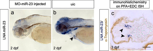
Specific miRNA expression in EDC fixed embryos which can be complemented with immunohistochemistry. (a,b) MiR-23 expression at 2 dpf in PFA+EDC fixed embryos. (a) Upon injection of a miR-23 MO, miR-23 expression was lost, while highly specific expression (arrowhead in b indicating the endocardial cushions) was detectable in non-injected control embryos (b). (c) Transversal section of an embryonic heart at 3 dpf fixed in PFA+EDC probed for miR-23 (arrowheads point to endocardial cushions) complemented with immunohistochemistry to detect tropomyosin (brown) in the myocardium. Scale bar represents 50 μm. |