
- Title
-
Distinct patterns of notochord mineralization in zebrafish coincide with the localization of Osteocalcin isoform 1 during early vertebral centra formation
- Authors
- Bensimon-Brito, A., Cardeira, J., Cancela, L., Huysseune, A., and Witten, E.
- Source
- Full text @ BMC Dev. Biol.
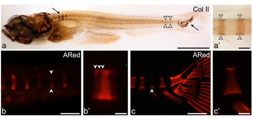
Early mineralization stages of vertebral centra and association with cartilaginous arches. Lateral view of (a, a′) Collagen type II immunostaining in the vertebral column of a 5.9 mm TL zebrafish shows (a) protein accumulation mostly in the cartilaginous structures of the Weberian apparatus and of the caudal fin skeleton (arrows) but also in the notochord sheath (white arrowheads). (a′) Although less evident than in cartilage, the staining can be clearly identified in the notochord matrix in a segmented manner (white arrowheads). (b-c) Early mineralization can be detected through alizarin red S (ARed) staining viewed under fluorescent light. (b) Early stage “ring” centrum (arrowheads) and (b′) close up showing the mineralization fronts (white arrowheads) in 5.0 mm TL fish. (c) Caudal fin centrum mineralization with a basiventral origin (arrowhead) and (c′) close up presenting a uniform mineralization surface with no distinct mineralization fronts, here represented by a 6.5 mm TL fish. Scale bars (a): 1 mm; (a′) 0.1 mm; (b,c): 0.15 mm; (b′, c′) 30 μm. EXPRESSION / LABELING:
|
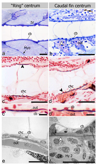
Chordacentrum formation and cell distribution inside and outside the notochord. Lateral view of 6.0 mm TL fish (a-d′). (a, b) Toluidine blue stained sections show the distinct morphologies of (a) “ring” and (b) caudal fin chordacentrum but homogenous chordoblast (cb) distribution during chordacentrum mineralization. In caudal fin vertebrae, outer (out) cells associated with the centra are chondrocytes from the cartilaginous arches (white arrowhead); (c and d) von Kossa staining highlighting that early (c) “ring” and (d) caudal fin centrum mineralization occurs within the notochord sheath, thus forming the chordacentrum (chc; black arrowheads); (c′ and d′) details showing outer and inner cells in both (c′) “ring” (dorsal side) and (d′) caudal fin centra (ventral side). Lateral view of (e) 5 mm TL fish in TEM micrograph showing that both chordoblasts and outer cells (out) display a squamous morphology, associated with sites of “ring” chordacentrum mineralization, while in caudal fin centra (f) the cartilaginous matrix of the arches is directly attached to the notochord sheath (white arrowhead), leaving no space for cells other than chondrocytes (cnc) to interact with the chordacentrum outside the notochord (11 mm TL fish). Further abbreviation: Hy2 – Hypural 2; IV – intervertebral space; Myo – myoseptum. Scale bars (a-d): 50 μm; (c′, d′): 10 μm; (e, f): 20 μm. |

utocentrum development and chordoblast positioning in the intervertebral space. Lateral view of (a) Toluidine blue stained section shows bone (bo) deposition around the chordacentrum (chc), forming the autocentrum in “ring” vertebrae. (b) TEM micrograph showing the intervertebral region (IV). Osteoblasts (ob) are clearly associated with bone deposition. (c) Autocentrum formation also follows chordacentrum development in caudal fin vertebrae although with apparent less amount of bone and osteoblasts due to the presence of the cartilaginous arches (white arrowheads). (d) TEM micrograph of the intervertebral (IV) region between two caudal fin centra showing the bone associated osteoblasts. (e) Sagittal section through the notochord at the level of the IV space. Once chordacentra are fully defined, as shown by von Kossa staining, chordoblasts (cb) tend to accumulate in the intervertebral region, acquiring an oblong shape, larger size, and positioning in a dorsal-ventral direction. (f) Sagittal section through the notochord at the level of the IV space. At this stage chordoblasts become highly proliferative, as confirmed by PCNA positive staining. Sections from panels a-d are from 11 mm TL fish, while e-f represent an earlier developmental stage with 6 mm and 6.2 mm TL fish, respectively. Further abbreviations: HPU2-3 - Haemal arch of Preural 2 and 3; Hy2 – Hypural 2; nsh – notochord sheath. Scale bars (a, c): 50 μm; (b, d): 10 μm; (e, f): 30 μm. EXPRESSION / LABELING:
|

Mineralization and centrum formation, lateral views, anterior left. (a) ALP in a “ring”-centrum (5.0mm TL) adjacent to chordacentrum (chc) mineralization site (arrowheads). (b) In caudal fin centrum (6.2mm TL) ALP (arrowhead) in arch-chondrocytes next to chordacentrum. Fast red counterstaining. Osteocalcin (arrowheads) in early mineralized (c) “ring”-centra (4mm TL), and (d) caudal fin centra (5.9mm TL). Osteocalcin in later stages: (e) “Ring”-centra (5.9mm TL), incremental marks (arrowheads), and (f) caudal fin centra, uniform staining. Sections of wholemount Osteocalcin immunostaining (g-j). In early chordacentra (5.9mm TL) Osteocalcin accumulates in the notochord sheath in: (g) “ring”-centra and (h) caudal fin centra. Co-localization with arch attachment (arrowhead). (g′) Detail of (g). Chordoblasts (cb), outer cells (out). (h′) Detail of (h). Osteocalcin-positive chondrocytes (out). In advanced chordacentra (i, j) uniform Osteocalcin accumulation in the matrix, 6.1mm and 6.9mm TL. (k) Tg(oc2:gfp; osx:mcherry) transgenic fish, 12mm TL. oc2-positive cells (green), at neural and haemal arches next to “ring”-centra (arrowheads). Osx-positive cells (red) cover vertebral centra. Gradient between (k′) less developed (oc2-negative) and (k′′) more developed (oc2-positive) centra. (k′′) Oc2 cells (white-arrowhead) next to an autocentrum. (l, m) qPCR, relative gene expression. oc1 and oc2. First detection of transcripts is the reference, represented by a 1-fold change (oc1:8-32 cells, oc2: 15dpf). Oc1, maternally transcribed, decreases at 12hpf (p<0.01), increases after 30dpf (p<0.01). Oc2, detected at 15dpf, increases at 45dpf (p<0.001). Age-size conversion under graphs. Hy1 – Hypural 1; IV – intervertebral space; n.d. – not detected. Scale bars: (a-b, g′-h′) 10 μm; (c-f, k) 1mm; (g-j, k′-k′′) 0.5mm. EXPRESSION / LABELING:
|