
- Title
-
Variation of BMP3 Contributes to Dog Breed Skull Diversity
- Authors
- Schoenebeck, J.J., Hutchinson, S.A., Byers, A., Beale, H.C., Carrington, B., Faden, D.L., Rimbault, M., Decker, B., Kidd, J.M., Sood, R., Boyko, A.R., Fondon, J.W., Wayne, R.K., Bustamante, C.D., Ciruna, B., and Ostrander, E.A.
- Source
- Full text @ PLoS Genet.
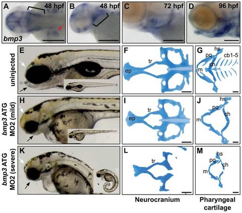
Zebrafish cranioskeletal development requires Bmp3 function. (A–H) Wholemount RNA in situ hybridization of bmp3 expression at 48 hpf (A,B), 72 hpf (C), and 96 hpf (D) stages. Anterior to the left. (A) Dorsal view, (B–D) lateral view. Pharyngeal arches indicated by brackets, pectoral fins by red arrowheads. Wholemounts (E,H,K) and alcian blue cartilage stains (F,G,I,J,L,M) of 96 hpf embryos from uninjected (E–G) and morpholino-injected embryos (h–j, mild phenotype, n = 72/177; k–m, severe phenotype, n = 83/177). Phenotypic severity is distinguished by tail curling (compare insets). Loss of jaw structures (black arrows) and frontal bossing (white arrowheads) is apparent in both classes of morphants. Cartilage is severely dysmorphic, hypoplastic, or absent following Bmp3 knockdown. Abbreviations correspond to ceratobranchial (cb), ceratohyal (ch), eythmoid plate (ep), hyosymplectic (hs), Meckel′s (m), palatoquadrate (pq), and trabeculae (tr) cartilages. |
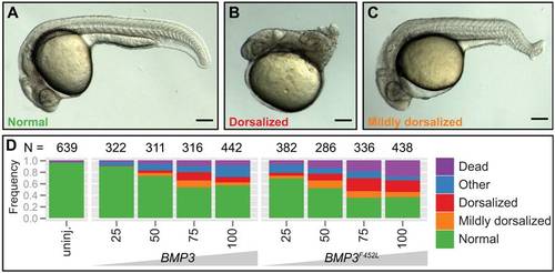
Overexpression activity differs between BMP3 variants. Overexpression utilized human BMP3 constructs, since the mature peptides of human and dog/wolf BMP3 are identical. (A–C) Whole mount embryos at embryonic stage 24 hpf, anterior to the left. Phenotypes are representative of (A) normal, (B) dorsalized, (C) mildly dorsalized classes following injection of human BMP3 mRNA into one-cell staged zebrafish embryos. (D) Stacked bar graph summarizing phenotypes observed following wt BMP3 or BMP3F452L mRNA injection. The dysmorphic phenotypes classified as “other” included combinations of mild dorsalization, tail curving, occlusion of the yolk extension, and invariably, hypoplasia or necrosis of head structures. Doses listed are in picograms (pg) of mRNA (x-axis). The frequencies of phenotypes are indicated by the y-axis. Each dose was repeated five or more times. The number of embryos injected is listed above each dose. Injection of BMP3F452L more potently dorsalizes embryos compared to wt BMP3 (student′s t-test P<0.05 for 25–75 pg doses, <0.01 for 100 pg dose). |
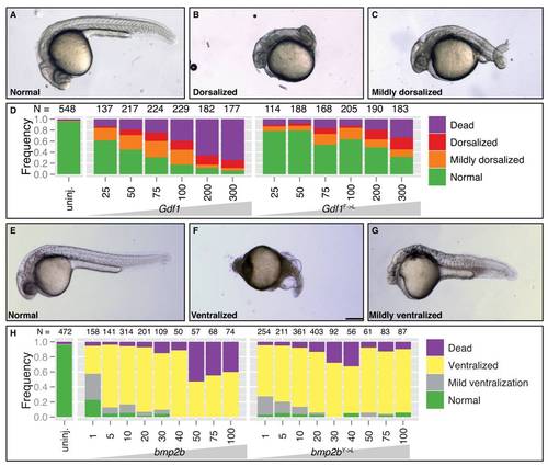
Y/F→L substitutions differentially affect Tgfßs. (A–C,E–G) Whole mount embryos at embryonic stage 24 hpf (A–C) or 28 hpf (E–G), anterior to the left. Phenotypes are representative of (A,E) normal, (B) dorsalized, (C) mildly dorsalized, (F) ventralized, (G) mildly ventralized classes following injections. (A–D) Embryos injected with mouse Gdf1or Gdf1F→L mRNA. (E–H) Embryos injected with either zebrafish bmp2b or bmp2bY→L mRNA. (D,H) Stacked bar graphs depicting frequency of observed phenotypes. Number of embryos injected per mRNA concentration appears above columns. While a missense mutation strongly reduces GDF1 dorsalizing activity, a comparable mutation in Bmp2b has little affect on this molecule′s ventralizing activity. |

Unillustrated author statements EXPRESSION / LABELING:
PHENOTYPE:
|