
- Title
-
Fibrillin-2b regulates endocardial morphogenesis in zebrafish
- Authors
- Mellman, K., Huisken, J., Dinsmore, C., Hoppe, C., and Stainier, D.Y.
- Source
- Full text @ Dev. Biol.

The scotch tape phenotype includes both a fin and a heart defect. Transmitted light images (A)-(D) show that the scote382 phenotype is visible by 24 hpf (A) and (B) and becomes clearer by 48 hpf (C) and (D). The caudal and dorsal fin fold defects manifest as a failure of the fin folds to expand, leading to a jagged appearance ((B), (D), black arrows) when compared to wild-type siblings (A) and (C). The heart defect can be accompanied by pronounced pericardial edema ((D), white arrow). Scale bars: (A) and (B), 0.5 mm; (C) and (D): 1 μm. |
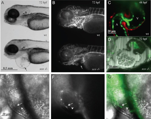
Heart vasculature defects in scote382 mutants. Fluorescence images of 72 hpf Tg(kdrl:GFP)s843 wild-type (A) and scote382 mutant (B) larvae. The overall patterning of the vasculature appears unaffected in scote382 mutant larvae; however, pronounced pericardial edema (black arrow in panel A) indicates a severe defect in heart function. Closer inspection reveals the absence of endocardial cells in the mutant atrium (white arrow in panel B). SPIM images show Tg(kdrl:GFP)s843; Tg(my17:DsRed)s879 wild-type endocardium and myocardium (C) at 48 hpf and the Tg(kdrl:GFP)s843scote382 mutant atrium, which appears to lack its endocardial lining (D) at 72 hpf. Panels C and D are stills taken from Video 1 and Video 2, respectively. Panels E–G are DIC images of a scote382 mutant atrium taken at 40×. An endocardial cell (white arrow) fails to adhere to its neighbor, thereby creating a gap (white arrowhead) in the endocardial sheet; DIC (E), the endothelial kdrl:GFP reporter (F) and merged image (G). A: atrium; V: ventricle. PHENOTYPE:
|

The endocardium fails to line the entire atrium in scote382 mutants. SPIM images of 48 hpf Tg(kdrl:GFP)s843; Tg(my17:DsRed)s879 embryos. Wild-type endocardium (A), (D) forms transient gaps, but recovers to line the entire chamber. A transient gap is outlined in white in panel D. The cardiac jelly (CJ) layer remains intact. scote382 mutant endocardial cells form transient gaps that develop into large areas devoid of endocardial cells (B), (C), (E) and (F). Endocardial gaps form in both the presence (A)–(C) and absence (D)–(F) of blood flow. Images taken in the presence of flow (A)–(C) are stills taken from movies documenting beating hearts ( Video 6). tnnt2 MO injected embryos (D)–(F) exhibit the same pattern of transient gaps in wild-type and scote382 mutants, with a stereotypical deficiency in the outer curvature of the atrium observed in mutants. A: atrium; V: ventricle. Scale bar: 20 μm. PHENOTYPE:
|

Endocardial cells form gaps during development, with a permanent deficiency manifesting in scote382 mutants. The mutant endocardium forms transient gaps, with a stable and permanent gap developing initially in the outer curvature of the atrium. Stills taken from a time-lapse SPIM movie (Movie S4) of one embryo document dynamic endocardial behavior in a tnnt2 MO injected kdrl:GFP scote382 mutant heart between 36 and 48 hpf. Frame number (n/361) is recorded in the bottom left corner of each panel. Red arrows point to transitory gaps, with gaps contributing to permanent holes outlined in red. In panels B, C and D, the larger outlined gap is in a proximal focal plane, with the smaller gap residing in the far wall of the atrium. Transitory gaps often form at the junction between three cells. Expected boundary of the atrial endocardial layer is indicated by the white dashed line. |

Electron microscopy study of endocardium details adhesion defects. Sections of 30 hpf wild-type (A), (C), (E) and scote382 mutants (B), (D), (F) at 9.4 kX (A) and (B) and 19 kX (C) and (D). Panels E and F correspond to the boxed areas of C and D, respectively. Wild-type endocardial cells (A), (C), (E) form a continuous layer between the cardiac jelly and the cardiac lumen. Mutant cells (B), (D), (F) fail to adhere to each other leading to a disorganized barrier, and allowing cardiac jelly to leak into the lumen. In (B), (D) and (F), the lumen and blood cells are located in the upper-left, while space between the endocardium and myocardium (which the cardiac jelly should occupy) is in the lower-right. Black arrows point to breaks between endocardial cells. CJ: cardiac jelly; BC: blood cell; L: lumen. PHENOTYPE:
|
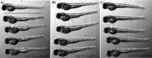
Complementation test between scote382 and pfdgw1. In a cross between scote382 heterozygotes and pfdgw1 heterozygotes, approximately 25% of embryos displayed a phenotype similar to that of scote382 mutants, including a blistered tail and pericardial edema. Panel A shows a scote382 incross, with a phenotypically wild-type embryo on top, and mutants below. Panel B shows the mutant embryos from a scote382 heterozygote to pfdgw1 heterozygote cross. Panel C shows phenotypically wild-type embryos from a scote382 heterozygote to pfdgw1 heterozygote cross. |
Reprinted from Developmental Biology, 372(1), Mellman, K., Huisken, J., Dinsmore, C., Hoppe, C., and Stainier, D.Y., Fibrillin-2b regulates endocardial morphogenesis in zebrafish, 111-119, Copyright (2012) with permission from Elsevier. Full text @ Dev. Biol.