
- Title
-
Macondo crude oil from the Deepwater Horizon oil spill disrupts specific developmental processes during zebrafish embryogenesis
- Authors
- de Soysa, T.Y., Ulrich, A., Friedrich, T., Pite, D., Compton, S.L., Ok, D., Bernardos, R.L., Downes, G.B., Hsieh, S., Stein, R., Lagdameo, M.C., Halvorsen, K., Kesich, L.R., and Barresi, M.J.
- Source
- Full text @ BMC Biol.

Exposure to Macondo crude oil-derived WAFs induced diverse gross morphological deformations in zebrafish embryos. (A-F) Lateral and ventral views of live untreated control (A, B) and WAF-treated embryos (C-F) at 5 dpf. Severe cardiac and yolk edema (C, D, E, arrows), dorsal tail curvature (E, F, double arrows), and cysts at the tip of the tail (C, D, F, arrowhead) were visible. (G, H) WAF-treated embryos (H) had reduced jaws compared to controls (G, brackets). (G-J) At 3 dpf cardiac edema was evident in WAF-treated embryos (arrows), and 28% of embryos had hemorrhaging in the forebrain, midbrain and hindbrain (arrowheads). Lateral (G, H, I, J) and dorsal views (H′, I′, J′). (K-M) Retinal architecture appeared normal in control and WAF-treated embryos (K, L) but there was a slight reduction in the area and perimeter of WAF-treated retinas (M). Except for lens area, the size reductions were statistically significant (M, asterisks; t-tests: lens area, P = 0.015; lens perimeter, P = 0.007; retina area P < 0.0005; retina perimeter, P < 0.0005). Scale bars: 200 μm, F, J′; 50 μm, L. Abbreviations: gcl, ganglion cell layer; ipl, inner plexiform layer; inl, inner nuclear layer; opl, outer plexiform layer; onl. outer nuclear layer. |

Macondo crude oil exposure did not affect cell proliferation but did induce programmed cell death. (A, B) Phospho-Histone H3 labeling of cells in mitosis were unaffected in 30 hpf WAF-treated embryos (B). (C) Quantification of anti-Activated Caspase 3-positive cells in 30 hpf control and WAF-treated embryos over 4 replicates and a WAF-refreshing procedure. The number of apoptotic cells decreased with each successive replicate, but increased following application of freshly-mixed WAF. (D-F) Activated caspase-3 labeled 30 hpf control (D, arrowheads) and WAF-treated embryos (E) from experiments in January 2011, and a WAF-treated embryo from an experiment in March 2011 (F, arrowheads). There was a significant decrease in the number of apoptotic cells in WAF-treated embryos between January (E) and March (F). (G-I) Representative images from the refreshed WAF experiments (arrowheads denote positive anti-Caspase 3 cells). Embryos were either untreated (G), exposed to the same WAF from 3.5 hpf to 30 hpf (H), or exposed to WAF from 3.5 hpf to 15 hpf and then exposed to a fresh WAF solution from 15 hpf to 30 hpf (I). Embryos in the refreshed WAF group (I) partially recovered the cell death phenotype of earlier replicates (E). (A, B, D-I) Lateral trunk views centered on somites 14 to 21. Scale bar 50 μm, A, B, D-I. |

Defects in head and trunk vascular development result in reduced circulatory function. (A-D) Hemoglobin staining revealed a reduction in the amount of blood cells in 3 dpf WAF-treated embryos, notably in the vasculature of the pharyngeal arches (A, B, brackets, lateral view; C, D, arrowheads, ventral view), with staining abruptly ending in the heart or bulbus artery prior to filling the aortic arches (arrow). (E-H′) Microangiography analysis with QTracker 655 fluorescent quantum dots (red) injected into 3 dpf tg[fli:eGfp] transgenic larvae to visualize endothelial cells associated with the vasculature (green). Endothelial vasculature in moderately affected WAF-treated embryos (G) was comparable to controls (E), however in severe cases posterior arch vasculature was lost and circulation was reduced (H, brackets, arrowhead, H′, arrowheads). (E′, F′, G′, H′) Arrowheads and arrows denote the specific blood vessels associated with the pharyngeal arches. Accumulation of quantum dots in the heart atrium suggests reduced flow into the ventricle (H′, dashed line). (I-K) Real time analysis of the flow speed of individual blood cells (I-J′, arrowheads) over a 7-somite distance in the dorsal aorta. WAF-treated embryos have reduced blood circulation (K, right half of graph). (L-O′) Intersegmental blood vessels had reduced circulation of quantum dots as demonstrated by either a complete absence of flow (M-O, arrowheads) or truncated flow (M-O arrows). Ectopic branching and vascular remodeling was evident in some segments devoid of circulation (N′, O′). Abbreviations: BuA, bulbus arteriosus; H, heart; HA, hypobranchial artery; ORA, opercular artery; PHS, primary head sinus. Numbers and affiliated arrowheads in E′ and F′ represent the first through sixth aortic arch. Scale bars = 200 μm, A-D; 100 μm, E-H′; 50 μm, I, J, N′, O′; 20 μm, I′, J′; 50 μm, L-O. |

Craniofacial defects induced by Macondo crude oil exposure were correlated with defects in neural crest development. (A-F) Alcian blue staining of head and jaw cartilage in 4 dpf control (A, D) and severely (B, E) or moderately (C, F) affected WAF-treated embryos. WAF-treated embryos had a variable reduction in the size of all cartilage components, notably a lack of anterior extension of jaw elements and a dramatic reduction in posterior pharyngeal arches (B, C, E, F). (G-L) Whole mount in situ hybridization of crestin expression in neural crest cells. crestin expression is normal in the trunks of control and WAF-treated embryos (G, I, K, bracket; circles in G, I, K represent magnified view in H, J, L). However, crestin expression was variably reduced specifically in the anterior migratory streams, an area of cells that will populate the pharyngeal arches (H, J, L, arrowheads and bracket). (M, N) Cranial neural crest forming pharyngeal arches (p1-5) at 31 hpf as visualized by fli driven expression of GFP. One of the posterior-most pharyngeal arches is missing in WAF treated embryos (N, arrows) as compared to controls (M, 3, 4, 5). (O, P) dlx2 expression in the region of pharyngeal arches is reduced in 26 hpf WAF-treated embryos (P, p1, arrow, bracket) as compared to controls (O). dlx2 expression is nearly lost in the most posterior regions of the presumptive pharyngeal arches (P, bracket) despite robust expression still seen in the forebrain. Abbreviations: bh, basihyal cartilage; cb1-5, ceratobranchial branches; ch, ceratohyal; m, Meckel′s; pq, palatoquadrate. Scale bars = 200 μm, A-F; 100 μm, G-L. |

Macondo crude Oil exposure impaired escape behavior by 48 hpf. (A) Individual frames from high-speed video recordings are shown for control larvae. The images are overlaid in 20 mS intervals and the duration of the response captured within the field is indicated. (B) Kinematics traces are shown for 10 escape responses each for control larvae. 0° indicates a strait body and positive and negative angles represent body bends in opposite directions. The time is indicated in seconds. (C) Image overlays for a WAF-treated larva escape response illustrates the failure to clear the field that was frequently observed. (D) Kinematic traces for WAF-treated larvae reveal reduced, abnormal body bend frequencies. (E) Quantification of body bend frequencies. (F) Quantification of the duration of escape responses reveals that WAF-treated larvae respond for shorter periods of time. Asterisks in E and F indicate statistically significant differences (n = 10, P < 0.01). |

Macondo crude oil exposure caused specific deformations in the peripheral but not central nervous system. (A-L) 30 hpf control and WAF-treated embryos labeled by immunohistochemistry. (A, B) 3A10 labeled Mauthner neurons in the hindbrain were normal. (C, D) Distribution of Gaba-positive interneurons in the spinal cord were not impacted by WAF treatment (d, DoLa; c, CoSa; v, VeLD; k, KA). (E, F) Anti-Acetylated tubulin (AT) labeling of primary motor axons (arrows) or Rohon Beard sensory neuronal somas (asterisk) were correctly positioned in WAF-treated embryos (lateral view of trunk and spinal cord). (G, H) Qualitatively, Islet1 labeling for primary and secondary motor neurons (lower bracket) and Rohon Beard sensory neurons (upper bracket) were positioned normally. (I, J) The branching pattern of AT-labeled sensory axonal projections along the trunk epidermis was significantly reduced (I′, J′, magnified views of boxed area in I, J). (K, L) Anti-Gfap labeled radial glia somas in WAF-treated embryo spinal cords (L) were correctly positioned in the ventricular zone (arrowheads) and similar in number to controls (K). (A, B) Dorsal views of the hindbrain. (C-L) Lateral views of the spinal cord (C, D, E, F, G, H, K, L) and trunk (I, J). Scale bars = 50 μm, A-L. |
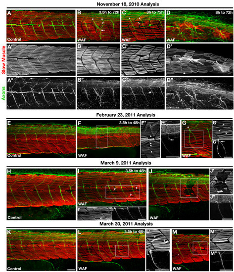
The severity of deformations in slow-twitch skeletal muscle development decreased with each experimental replicate. (A-M) Lateral views of F59 labeled Myosin heavy chains in slow-twitch muscle fibers (red) and anti-Acetylated tubulin labeled motor axons (green) in the embryonic trunk at 72 hpf (A-D) and 48 hpf (E-M). (A-D) Initial Macondo crude oil WAF treatments beginning at either 3.5 hpf (B) or 8 hpf (C, D) and ending at 72 hpf showed severely defective neuromuscular phenotypes, such as improper somite boundary formation (B, C, arrowhead) or slow muscle loss and disorganization (D). Somite boundary defects associated with the middle or ventral portion of the somite correlated with motor axon pathfinding errors (B, asterisks, arrow; D). (E-M) Subsequent WAF treatments beginning at 3.5 hpf and ending at 48 hpf induced somitogenesis (F, G; F, G′, arrowheads), slow muscle (I-M′, arrowheads), and motor axon pathfinding (F, G; F′′, G′′, arrows) defects, however the severity of these defects decreased over time with each experimental replicate from November to March. (A-M) Primed letters represent single channel images of slow muscle (single prime) or axon (double prime) labeling for the whole image (A-D) or just the boxed regions (F, G, I, J, L, M). Scale bars = 50 μm, A-M′′. |

Repeated application of freshly mixed WAF reproduced the severe skeletal muscle phenotypes. (A-E) Lateral views of the trunk of an untreated control embryo (A), WAF-treated embryos from 3.5 hpf to 48 hpf (B, C), and embryos exposed to freshly mixed WAF every 15 h (D, E). WAF-treated controls showed mild slow muscle defects (B′, C′), while embryos treated repeatedly with refreshed WAF displayed severe somite and slow muscle phenotypes (D′, E′). As seen in earlier experiments, slow muscle fibers spanned presumptive boundaries (D′, arrowheads), somitic shape was irregular (D′, arrows), and slow muscle degeneration was evident (E, a representative degenerating myofibril is pseudo-colored red). Scale bars = 50 μm, A-E′. |