
- Title
-
Smyd1b_tv1, a Key Regulator of Sarcomere Assembly, Is Localized on the M-Line of Skeletal Muscle Fibers
- Authors
- Li, H., Xu, J., Bian, Y.H., Rotllant, P., Shen, T., Chu, W., Zhang, J., Schneider, M., and Du, S.J.
- Source
- Full text @ PLoS One

Smyd1b_tv1myc and Smyd1b_tv2myc show dynamic localizations during muscle cell differentiation in zebrafish embryos. A–D. Whole-mount immunostaining with anti-myc antibody shows the cytosolic localization of Smyd1b_tv1myc (A, C) or Smyd1b_tv2myc (B, D) in myoblasts of transgenic zebrafish embryos at 14 and 24 hours-post-fertilization (hpf), respectively. A and B, dorsal views. C and D, side views. E, G, I, K. Immunostaining with anti-myc antibody shows the sarcomeric localization of Smyd1b_tv1myc in myofibers of transgenic fish embryos at 27, 30, 48, and 72 hpf, respectively. F, H, J, L. Immunostaining with anti-myc antibody shows the cytosolic (F, H, J) and sarcomeric (L) localization of Smyd1b_tv2myc in myofibers of transgenic fish embryos at 27, 30, 48, and 72 hpf, respectively. Scale bars: 30 μm. |

The sarcomeric localization of Smyd1b_tv1 occurs after the sarcomere formation in myofibers of zebrafish embryos. A–C. Immunostaining using sarcomeric specific antibodies against MyHC (A), α-actin (B), and α-actinin (C) in the trunk muscles of zebrafish embryos at 24 hpf. D. Immunostaining using anti-myc antibody shows the primary cytoplasmic localization of Smyd1b_tv1 in the trunk muscles of smyd1b_tv1myc transgenic fish embryos at 24 hpf. Muscle pioneer cells with the sarcomeric localization are indicated by arrows. Scale bar: 30 μm. |

Rescue of myofibril organization defect in smyd1b knockdown embryos by expression of Smyd1b_tv1-EGFP or Smyd1b_tv2-EGFP fusion protein. A. DNA constructs encoding Smyd1b_tv1-EGFP, or Smyd1b_tv2-EGFP fusion proteins or EGFP control were generated and injected into zebrafish embryos. B and C. Myofibers expressing Smyd1_tv1-EGFP (B) or Smyd1_tv2-EGFP (C) was directly observed by GFP. D and E. Myosin thick filaments organization was determined by F59 antibody staining in Smyd1_tv1-EGFP (D) or Smyd1_tv2-EGFP (E) co-injected embryos. F and G. Double staining shows the colocalization of normal fibers with Smyd1_tv1-EGFP (F) or Smyd1_tv2-EGFP (G) expression. Scale bars: 20 μm. |
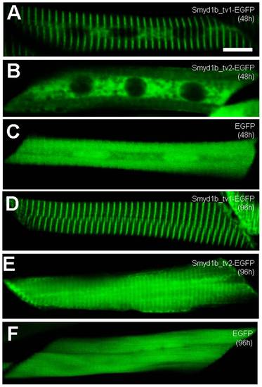
Characterization of the sarcomeric localization using Smyd1b_tv1-EGFP and Smyd1b_tv2-EGFP fusion proteins. DNA constructs encoding Smyd1b_tv1-EGFP, or Smyd1b_tv2-EGFP fusion proteins or EGFP control injected into zebrafish embryos. Their expression and localization was determined in myofibers of the injected zebrafish embryos at 48 (A–C) and 96 (D–F) hpf. A and D, Smyd1b_tv1-EGFP; B and E, Smyd1b_tv2-EGFP; C and F, EGFP control. Scale bar: 8 μm. |

Smyd1b_tv1-EGFP is localized on the M-line of zebrafish skeletal muscles. Smyd1b_tv1-EGFP construct was injected into Myomesin-RFP (A, C, E) or wild type (B, D, F) zebrafish embryos at 1–2 cell stages. Smyd1b_tv1-EGFP localization was determined together with M-line marker (Myomesin-RFP) and A-band marker (Myosin heavy chain) at 96 hpf. A, B and C. Co-localization of Smyd1b_tv1-EGFP and Myomesin-RFP was observed in myofibers of the injected embryos. D, E and F. Immunostaining with anti-MyHC antibody (F59) shows the localization of Smyd1b_tv1-EGFP in the middle of the A-bands in myofibers of zebrafish embryos. Scale bar: 12 μm. |

Smyd1b_tv1myc is localized on the M-line of adult zebrafish skeletal muscles. A and B. Immunostaining using anti-myc antibody shows the sarcomeric localization of Smyd1b_tv1myc on longitudinal sections of skeletal muscles from adult transgenic zebrafish expressing a myc-tagged Smyd1b_tv1 (A) or non-transgenic control (B). C–E. Double immunostaining with anti-myomesin and anti-myc antibodies shows the colocalization of Smy1b_tv1myc with myomesin on the M-lines. F–H. Double immunostaining with anti-α-actinin and anti-myc antibodies shows the non-overlapping localization of Smy1b_tv1myc with α- actinin. Scale bars: A = 22 μm. C = 6 μm. |
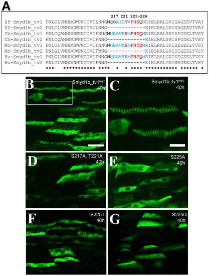
The Serine 225 is required for the enhanced sarcomeric localization of Smyd1b_tv1. A. Sequence comparison shows that the alternative splicing of smyd1b in various vertebrates and the conserved serine and threonine residues within the 13 aa insertion. B and C. Immunostaining using anti-myc antibody shows the sarcomeric localization of Smyd1b_tv1myc in myofibers of zebrafish embryos at 38 hpf. C represents the highlighted box area in A. D–G. Immunostaining using anti-myc antibody shows the sarcomeric localization of Smyd1b_tv1myc mutant proteins that carry substitutions at S217A and T221A (D), S225A (E), S225T (F), S225D (G). Scale bars: B = 40 μm; C = 20 μm. |
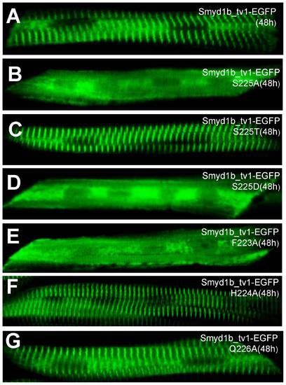
Effect of S225A, S225T, S225D, F223A, H224A and Q226A substitution on the sarcomeric localization of Smyd1_tv1-EGFP in zebrafish embryos. DNA construct expressing Smyd1_tv1-EGFP or its derived mutants of S225A, S225T, S225D, F223A, H224A and Q226A was injected into zebrafish embryos. Their localization was analyzed in myofibers of the injected embryos at 48 hpf. A, Smyd1_tv1-EGFP; B, Smyd1_tv1-EGFP(S225A); C, Smyd1_tv1-EGFP(S225T); D, Smyd1_tv1-EGFP(S225D), E, Smyd1_tv1-EGFP(F223A), F, Smyd1_tv1-EGFP(H224A), and Smyd1_tv1-EGFP(Q226A). |
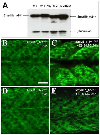
Knockdown of endogenous Smyd1b advances the timing of sarcomeric localization of Smyd1b_tv1myc in zebrafish embryos. A. Smyd1b E9I9-MO was injected into Smyd1b_tv1myc or Smyd1b_tv1myc transgenic zebrafish embryos at 1–2 cell stages. Western blot analysis shows the expression of myc-tagged Smyd1b_tv1myc and Smyd1b_tv1myc in un-injected control or E9I9-MO injected transgenic zebrafish embryos at 24 hpf. γ-Tubulin was used as loading control. B and C. Immunostaining using anti-myc antibody shows the cytoplasmic (B) or sarcomeric localization (C) of smyd1b_tv1myc in control (B) or E9I9-MO injected (C) transgenic zebrafish embryos at 24 hpf. D and E. Immunostaining using anti-myc antibody shows the cytoplasmic localization of smyd1b_tv2myc in control or E9I9-MO injected transgenic zebrafish embryos at 24 hpf. Scale bar: 30 μm. |
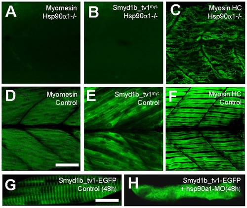
The effect of hsp90α1 mutation or knockdown on Smyd1b_tv1 sarcomeric localization. A and D. Immunostaining using anti-myomesin antibody shows the organization of myomesin in hsp90α1 mutant (A), or control (D) smyd1b_tv1myc transgenic embryos at 72 hpf. B and E. Immunostaining using anti-myc antibody shows the localization of Smyd1b_tv1myc in hsp90α1 mutant (B), or control (E) smyd1b_tv1myc transgenic embryos at 72 hpf. C and F. F59 staining shows the organization of slow muscle myosin in hsp90α1 mutant (C), or control (F) smyd1b_tv1myc transgenic embryos at 72 hpf. G and H. DNA construct Smyd1_tv1-EGFP was injected alone or together with hsp90α1 ATG-MO into zebrafish embryos. Smyd1_tv1-EGFP localization was determined in myofibers of the control (G), or hsp90α1 knockdown (H) at 48 hpf. Scale bars: D = 40 μm; G = 15 μm. |