
- Title
-
Qilin is essential for cilia assembly and normal kidney development in zebrafish
- Authors
- Li, J., and Sun, Z.
- Source
- Full text @ PLoS One

qilinhi3959A mutant develops body curvature and kidney cysts. (A, B) Side view of embryos at 3 dpf. Inset in B is a zoomed in view of the kidney cyst in the mutant. (C–D) Cross section through the glomerular-tubular region of embryos at 5 dpf. Red arrow in C points to the fused glomeruli, while red dotted lines in D outline the cysts. (E–F) Cross sections of the duct of embryos at 5 dpf. Solid green lines outline the duct. Green arrows point to the duct. (G–H) Side view of the pronephric duct in whole-mount embryos at 5 dpf stained with Cdh-17. Yellow dotted lines outline the duct. WT: wild type; MUT: mutant. Scale bars in C–F: 20 μm, in G and H; 5 μm. |

hi3959A is a zygotic null allele of qilin. (A) Graphic representation of qilin gene. Blue squares represent exons. Red triangle indicates the proviral insertion site. Arrows indicate the direction and location of the genotyping primers used. (B) qilin transcript in embryos at 5 dpf shown by RT-PCR of cDNA from hi3959A embryos (lane 2) and wild type siblings (lane 1). Lane 3 and 4 are beta-actin loading controls for hi3959A (Lane 4) and wild type siblings (Lane 3). (C) Genotyping PCR of embryos from hi3959A carrier in-crosses injected with eGFP-qilin mRNA. All embryos genotyped exhibited wild type phenotypes. Lower band is specific for the mutant allele, while the upper band is specific for the wild type allele. From left to right, the three embryos are heterozygous, homozygous mutant and homozygous wild type, respectively. (D) Graph displaying percent embryos with the curved body phenotype in hi3959A carrier-in crosses that are uninjected (n = 3, with an average of 140 embryos per experiment) and injected with eGFP-qilin mRNA (n = 3, with an average of 120 embryos per experiment). * p< 0.05. EXPRESSION / LABELING:
|

qilin is expressed maternally and ubiquitously. (A–F) In situ hybridization for qilin on embryos at the 8- cell stage (A), the shield stage (B), the 3–5 somites stage (C), and the 24 hours-post-fertilization (hpf) stage (E). D and F are sense controls at the 3–5 somites stage (D) and the 24 hpf stage (F). (G) qilin transcript shown by RT-PCR from 16 cell (Lane 1) and 2 dpf (Lane 2) wild type embryos. Lane 3 and 4 are elf1a loading controls for 16-cell (Lane 3) and 2 dpf (Lane 4) samples with matching dilutions of cDNA used in PCR reactions. |

Pronephric cilia in qilin hi3959A mutants are defective. (A–D) Epifluorescent projections showing the pronephric cilia in a wild-type sibling (A, C) and a mutant (B, D) at the 1 dpf; in both anterior (A, B) and posterior (C,D) portions of the duct. Yellow arrowheads in A and B point to rows of basal bodies in MCCs. (E, F) Epifluorescent projections showing the pronephric cilia in a wild-type embryo (E) and a mutant (F) at 5 dpf in the posterior portion of the duct. All embryos were stained with anti-γ-tubulin (red), anti-Sco (green), and DAPI (blue). EXPRESSION / LABELING:
PHENOTYPE:
|

Single cilia form earlier in development than multi-cilia in the pronephric duct. (A) Graphical representation of multi-cilia observed in embryos at 24 somite, 24 hpf, and 30 hpf. Single cilia were observed at all time points analyzed. Bars represent percentage of embryos that developed multi cilia in the pronephric ducts. Each bar represents data from three independent experiments with at least 8 embryos each. (B–D) Representative images of cilia at 24-somite (B), 24 hpf (C) and 30 hpf (D). Yellow arrowheads point to basal bodies of multi-cilia. (E) A model of how different developmental timing of single cilia and multi-cilia could contribute to the pronephric cilia phenotypes observed in hi3959A mutants. |
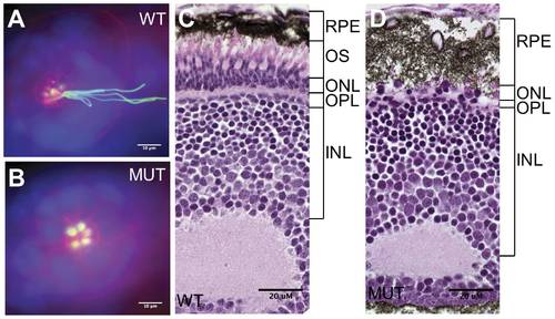
Sensory cilia in qilin hi3959A mutants are defective. (A–B) Epifluorescent projections showing the lateral line organ in a wild type (A) and a mutant embryo (B) at 3 dpf. Embryos were stained with rodamine-phalloidin (red), anti- acetylated tubulin (green) and DAPI (blue). Scale bars: 10 μm. (C–D) The absence of the outer segment of photoreceptors in the eye as shown through cross sections of a wild type (C) and a mutant embryo (D) at 5 dpf. WT: wild type; MUT: mutant; RPE: retinal pigment epithelium; OS: outer segment; ONL: the outer nuclear layer; INL: the inner nuclear layer; OPL: the outer plexiform layer. Scale bars: 20 μm. PHENOTYPE:
|
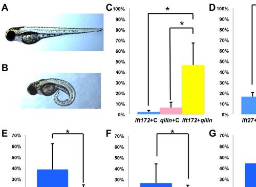
qilin genetically interacts with ift172 and ift27. (A–B) Side view of a representative uninjected control embryo (A) and a phenotypic embryo injected with qilin and/or ift172, ift27 morpholino (B) at 2 dpf. (C, D) Graph displaying the percent of embryos that develop curved bodies. In C, embryos are either injected with qilin morpholino and the control morpholino; ift172 morpholino and the control morpholino; or qilin morpholino with ift172 morpholino. In D, embryos are either injected with qilin morpholino and the ift27 mismatched-control morpholino; ift27 morpholino and the ift27 mismatched-control morpholino; or qilin morpholino with ift27 morpholino. (E–G)Graphical representation of the effectiveness of ift27:GFP in rescuing body curvature (E), kidney cysts (F), and laterality defects (G) observed in ift27 morphants. Embryos are either injected with ift27 morpholino and myc:GFP mRNA (blue bars), or ift27 morpholino with ift27:GFP mRNA (pink bars). Laterality defects in (G) is presented as the percentage of embryos developed hearts positioned on the right or center. N = 3 for all experiments, with at least 40 embryos per experiment per condition. *: p< 0.05, **: p< 0.01. MO: morpholino. PHENOTYPE:
|

Unillustrated author statements |