
- Title
-
Evolutionary relationships and diversification of barhl genes within retinal cell lineages
- Authors
- Schuhmacher, L.N., Albadri, S., Ramialison, M., and Poggi, L.
- Source
- Full text @ BMC Evol. Biol.
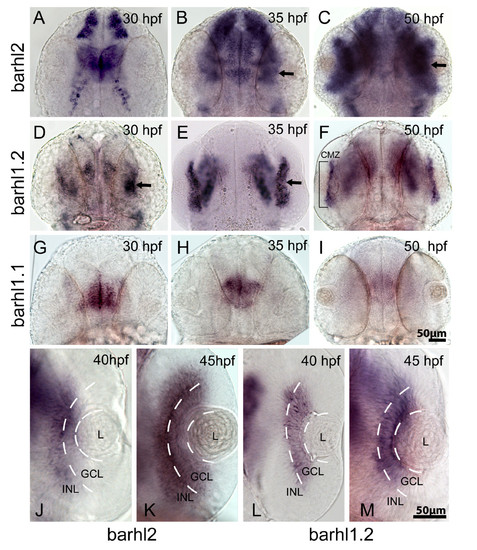
Comparative in situ hybridization of barhl paralogs expression in the zebrafish retina. Dorsal view of wild-type zebrafish embryos hybridized with barhl2 (A-C, J-K), barhl1.2 (D-F, L, M) and barhl1.1 (G-I), antisense RNA probes. Stages analyzed are indicated. Anterior is always to the top. (B, C, D and E) black arrows in indicate expression localized in the retina. In (F), the black bracket highlights barhl1.2 expression restricted in a thin retinal domain, which is the ciliary marginal zone (CMZ). (J-M) show a closer view on individual retinas. White dashed lines highlight the boundary between lens (L), ganglion cell layer (GCL) and inner part of the inner nuclear layer (INL), where ACs are located. |
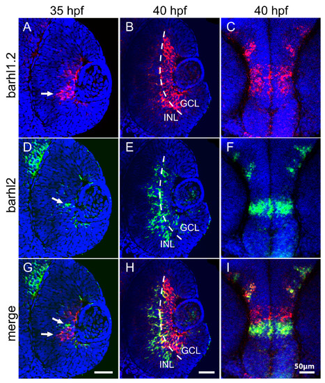
Double fluorescent in situ hybridization of barhl1.2 and barhl2. Confocal sections through the central retina (A-B, D-E and G-H) or diencephalon (C, F and I) of embryos hybridized with both barhl1.2 and barhl2 antisense RNA probes. Stages analyzed are indicated. Nuclei were stained with DAPI (blue). All pictures represent a frontal view (anterior is always to the top). (A-C) barhl1.2 RNA antisense probe revealed with Cy3 (red). (D-F) barhl2 RNA antisense probe revealed with FITC (green). (G-I) green and red channel merged. White arrows in (A, D and G) indicate non-overlapping expression of the two genes. Dashed line in (B, E and H) highlights the boundary between the ganglion cell layer (GCL) and the inner nuclear layer (INL). |
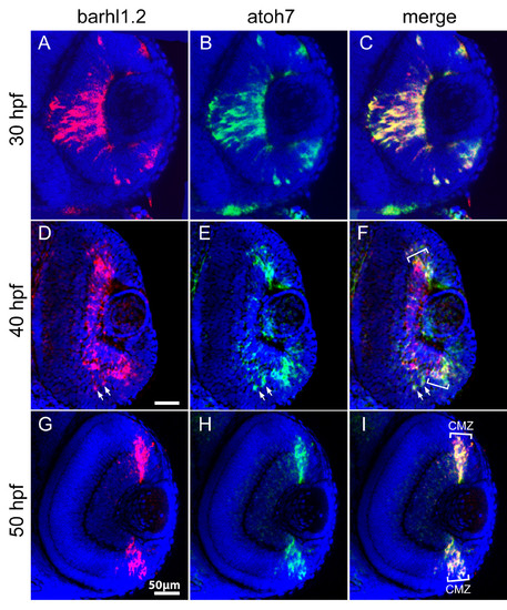
Double fluorescent in situ hybridization of barhl1.2 and atoh7. Confocal sections through the central retina of embryos hybridized with barhl1.2 (A, D and G, in red) and atoh7 (B, E and H, in green) antisense RNA probes. (C, F and I) merge of red and green channels. Nuclei were stained with DAPI (blue). View is frontal in all pictures, anterior is always to the top. Stages analyzed are indicated. (D- 25 -F) Downregulation of barhl1.2 in the central retina is delayed with respect to the one of atoh7 but overlapping in the ciliary marginal zone (highlighted with white brackets CMZ). White arrows highlight two cells where both barhl1.2 and atoh7 are expressed. (F and I) the white brackets indicate the CMZ where barhl1.2 and atoh7 expression always overlap. |
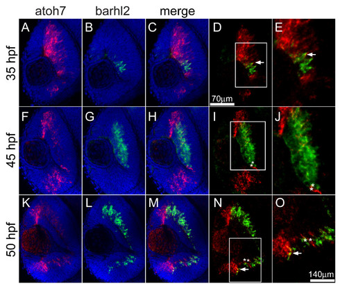
Double fluorescent in situ hybridization of barhl2 and atoh7. Confocal sections through the central retina of embryos hybridized with barhl2 (revealed with FITC, shown in green) and atoh7 (revealed with Cy3, shown in red), antisense RNA probes. Nuclei were stained with DAPI (blue) to outline retinal layers. View is frontal in all pictures, anterior is always to the top. (D-E and N-O) White arrows show co-localization of both mRNAs in cells at the border of the expression domains. (I-J and N-O) white asterisks indicate adjacent cells expressing either barhl2 or atoh7. (D, I and N) white squares highlight the magnified area in E, J and O, respectively. |

Expression of barhl paralogs in the atoh7-/- retina. 40 hpf zebrafish embryos hybridized with barhl2 (A-D) or barhl1.2 (E-H) RNA antisense probe. (A, E and B, F) show dorsal view of wt (A, E) and atoh7-/- mutant (B, F) retinas, anterior is to the top. barhl1.2 expression is absent in the atoh7-/- retina (F). (C, D) dorsal view and (G, H) lateral view at the level of the tectum (t) and rhombic lips (rl), showing that expression of both barhl2 (C, D) and barhl1.2 (G, H) remains unchanged in these brain areas. White arrow in G indicates barhl1.2 expression in the retina, which is missing in atoh7-/- mutants (H). |

Expression patterns of the medaka barhl1 and barhl2 in the retina. Dorsal views through the retina of medaka embryos hybridized with either medaka barhl1 (A, B) or medaka barhl2 (C, D) RNA antisense probe. Anterior is to the top. (A) Expression of medaka barhl1 restricted to the ciliary marginal zone (CMZ) is highlighted with black brackets in A and with a black dotted line in B. The white dotted line indicates the ganglion cell layer (GCL)/inner nuclear layer (INL) boundary. |

Unillustrated author statements EXPRESSION / LABELING:
|