
- Title
-
Induction of apoptosis and inhibition of cell growth by tbx5 knockdown contribute to dysmorphogenesis in Zebrafish embryos
- Authors
- Lu, J., Tsai, T., Choo, S., Yeh, S., Tang, R., Yang, A., Lee, H., and Lu, J.
- Source
- Full text @ J. Biomed. Sci.
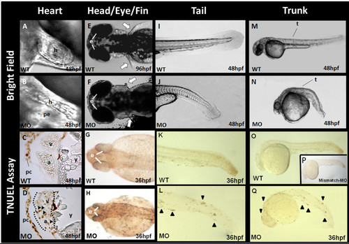
Morphological changes and the TUNEL assay of tbx5 knockdown embryos. A: In wild-type (WT) embryos, the atrium and ventricle overlap due to normal looping formation. The heart is located in the cardiac sac. B: In tbx5 knockdown embryos, a string-like cardiac morphology was observed, with no looping formation. Most of the cardiac sac was occupied by pericardial effusion. C: No apoptosis was observed in the atria or ventricle of WT embryos, but a little apoptosis was noted in the pericardium. D: Apoptosis occurred in the heart of tbx5 knockdown embryos accompanied by significant apoptosis in the pericardium. E-F: Symmetrical development of bilateral pectoral fins was observed in WT embryos (E); however, pectoral fins of tbx5 deficient embryos were hypoplasia (F). G: Trivial apoptosis was observed in the eyes, head, and the bilateral paravertebral mesenchymal region of WT embryos. H-J: Prominent apoptosis was detected in knockdown embryos (H). Compared to WT embryos (I, M), a shortened and curled malformed trunk was present in tbx5 knockdown embryos (J, N). K-Q: Prominent apoptosis was located along the curled tail (L) and trunk (Q) of tbx5 deficiency embryos, while normal embryos and mismatch-tbx5-MO injected embryos had a straight tail (K) and trunk (O, P). A-Q: The anterior of the embryos is to the left. A-B, I-Q: Lateral view; E-F: dorsal view; G-H: anterior view; C, D: Sagittal section. h, heart; pe, pericardial effusion; a, atrium; v, ventricle; y, yolk; t, trunk; white arrow, pectoral fin; black triangle, location of apoptosis; hpf, hour post fertilization; MO, tbx5-MO treated embryos. |

Quantification of apoptosis-related gene expressions in tbx5 deficiency zebrafish embryos using a semi-quantitative PCR. A: The efficiency of the tbx5-MO was tested using Western blot. Tbx5-MO remarkably inhibited the expression of the TBX5 protein (n = 3, 100 embryos). B-C: The proapoptotic genes, bad (B) and bax (C), were significantly activated in tbx5-MO-treated embryos. D: The antiapoptosis gene, bcl2, was significantly induced in tbx5 knockdown embryos throughout the developmental stages (n = 3, 50 embryos/stage; relative expression, gene expression/²-actin expression). Data are presented as the mean ± S.D. An asterisk indicates a significant difference (p < 0.05). |
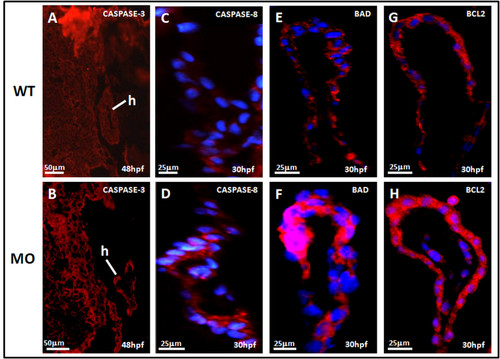
Sagittal view of the heart in tbx5 knockdown embryos by immunohistochemical studies of heart. A-B: Caspase-3 (red) expression could been slightly seen in wild-type (WT) embryos (A), but caspase-3 was more induced in knockdown embryos at 48 h post-fertilization (hpf) though the expression was not so strong (B). Caspase-8 (blue) was more induced in knockdown embryos (D) at 48 h post-fertilization (hpf) than the expression in wild-type (C). The proapoptotic factor, BAD (red), was enhanced in tbx5 deficiency embryos (F) with the nucleus stained by DAPI (blue); but there was almost no BAD expression in WT embryos (E). Compared to the WT (G), embryos with tbx5 deficiency showed a significant increase in BLC2 expression (H). The anterior of embryos is to the left. h, heart; MO, tbx5-MO-treated embryos. EXPRESSION / LABELING:
|

Immunohistochemical studies of heart sagittal sections in tbx5 knockdown embryos by cell cycle-related antibodies. A-D: Both cell cycle-related factor CDK2 (red) (A) and P27 (red) (C) expressed mildly in WT embryos but was highly induced in tbx5 knockdown embryos (B, D) at 30 hpf. The nucleus was counterstained with DAPI. The anterior of embryos is to the left. h, heart; MO, tbx5-MO-treated embryos. |

Myosin of zebrafish embryos was stained by the heart-specific anti-myosin antibody, MF20 (green), and counterstained by DAPI (blue) for nuclear observation. A-F: The expression of MF20 in embryos with tbx5 deficiency (B, D, F) decreased throughout the developmental stages compared to that in the WT (A, C, E), from 24 to 48 hpf. The anterior of embryos is to the left. h, heart; MO, tbx5-MO-treated embryos. |

Ultrastructural changes of mitochondria and myofibrils in the heart of tbx5-knockdown and wild-type (WT) embryos at 48 h post-fertilization (hpf) under TEM. A: Small mitochondria and well-formed desmosomes can be observed in WT embryos. B: Multiple swollen mitochondria can be observed within myocytes of tbx5 knockdown embryos. C: Well-organized myofibrils with a M line and Z disc can be observed in normal embryos. D: Disarrayed myofibrils appeared in tbx5 knockdown embryos while the M line and Z disc were unrecognizable. E-F: Both the size (E) and number (F) of mitochondria had significantly increased in tbx5 knockdown embryos. Data are presented as the mean ± S.D. An asterisk indicates a significant difference (p < 0.05). mf, myofibril; mt, mitochondria; D, desmosome; ML, M-line; Z, Z-disc; white arrow, disarrayed myofibrils; MO, tbx5 knockdown embryos. PHENOTYPE:
|