
- Title
-
Gal80 intersectional regulation of cell-type specific expression in vertebrates
- Authors
- Fujimoto, E., Gaynes, B., Brimley, C.J., Chien, C.B., and Bonkowsky, J.L.
- Source
- Full text @ Dev. Dyn.

Pan-neuronal expression of Gal80 does not affect central nervous system (CNS) development. Whole-mount embryos (transgenic Tg(elavl3:Gal80)zc64 or nontransgenic sibling), ventral views (except D), anterior to the top. Images are confocal projections (except A and B). A–A′: Brightfield images showing expression of gal80 by in situ in wild-type (A) and transgenic embryos (A′). B–B′: dlx2 in situ expression in wild-type and transgenic embryos is similar. C–C′: Pattern of tyrosine hydroxylase (TH) antibody expression is similar in wild-type and transgenic embryos. D–D′: Axon tract architecture appears similar in wild-type and transgenic embryos (lateral views, anterior to the left). E–E′: Acridine orange staining for apoptotic cells is similar in wild-type and transgenic embryos. Scale bar = 50 μm. |

Gal4 drives strong expression in stable transgenic lines and can be inhibited by Gal80. Confocal maximum projections of 72 hours postfertilization (hpf) embryos; ventral views, anterior to the top. Embryos are heterozygous for the genotypes shown. Arrow points to ectopic green fluorescent protein (GFP) expression in eyes driven by Gal4-VP16 (A,D) not noted using native Gal4 (E). A–C: Live embryos imaged with identical laser power settings, showing that Gal4-driven expression (B) is stronger than a direct enhancer:GFP construct (C) and is comparable to Gal4-VP16-driven expression (A). D–F: Immunohistochemistry for GFP in fixed embryos again shows that Gal4-dependent expression (E) is comparable to Gal4-VP16 (D), but has minimal expression in other tissues (such as eye muscles, arrow). F: The original transgenic enhancer line Tg(otpb.A:egfp)zc48 shows dimmer expression compared with Gal4-driven expression in E. G: A weakly expressing allele of elavl3:Gal80 only partially inhibits Gal4-dependent transgene expression. H: A strongly expressing elavl3:Gal80 allele completely inhibits Gal4-dependent expression. Insets for G and H show relative levels of Gal80 in situ expression. I: Gal80 is unable to inhibit Gal4-VP16-driven expression. Scale bar = 50μm. |

Time course of Gal80-mediated inhibition of Gal4. Confocal images of live embryo green fluorescent protein (GFP) expression. Transgenic fish carrying Tg(hs:Gal80); Tg(otpb.A:Gal4); Tg(UAS:GFP) were imaged before a 452 heat-shock, and then at defined time intervals following heat-shock. The same embryo is displayed in each respective set of panels (A–A′′ ′′ or B–B′′ ′′). Arrow points to the inhibition of Gal4-dependent expression in (A′); arrowhead points to the return of GFP expression in (A′′ ′′) 24 hr following the end of heat-shock. Images are maximum projections of 10 z-slices. Ventral views, rostral to the top, identical confocal settings for imaging both embryos. Scale bar = 50 μm; “h”, GFP expression in the heart from the transgenesis marker. |

Temporal control of Gal4-dependent expression using a temperature sensitive version of Gal80. Confocal maximum intensity projections, ventral views, anterior to top, 72 hours postfertilization (hpf) Tg(otpb.A:Gal4)zc67; Tg(UAS:GFP) embryos stained with anti-green fluorescent protein (GFP). A,B: Expression is indistinguishable between embryos without (A) or with (B) the Tg(elavl3:Gal80ts)zc68 transgene when raised at the restrictive temperature (28.5°C). C,D: When raised at permissive temperatures (21.5°C or 23°C) starting at 24 hpf, Gal80ts is able to effectively inhibit Gal4-dependent expression. E: When Gal4-dependent expression is initially allowed to occur from 24 to 48 hpf, shifting to the permissive temperature from 48–72 hpf allows Gal80ts to inhibit Gal4-dependent expression. F: Raising at a permissive temperature until 48 hpf, then relieving Gal80-mediated inhibition at 48 hpf permits Gal4-dependent expression to resume by 72 hpf. G,G′: Live confocal images of the same embryo before (at 24 hpf) and after (at 36 hpf) shifting to the permissive temperature, shows inhibition of Gal4-dependent expression. H: In comparison, a control embryo raised at the restrictive temperature only shows robust expression. Inset shows this same embryo at 24 hpf. Scale bar = 50 μm. |

Nuclear localization and codon optimization improves function of Gal80. Confocal maximum projections, lateral views, anterior to left, dorsal up, of eyes in Tg(isl2b.3:Gal4)zc65; Tg(UAS:GFP) 72 hours postfertilization (hpf) embryos. Immunostaining for green fluorescent protein (GFP), green; Topro3 nuclear stain, magenta. A–A′′ ′: Tg(isl2b.3:Gal4)zc65; Tg(UAS:GFP) transgenic embryos with no Gal80 show GFP expression in all retinal ganglion cells (RGCs). Inset and A′′ ′ shows high power magnification of single confocal slice. B–B′′ ′: Triple transgenic Tg(isl2b.3:Gal4)zc65; Tg(UAS:GFP); Tg(brn3c:Gal80) shows inhibition of Gal4-dependent GFP expression in approximately 30% of RGCs. Inset and B′′ ′ shows high power magnification of single confocal slice. C–C′′ ′: Transient injection with construct carrying “improved” Gal80 into Tg(isl2b.3:Gal4)zc65; Tg(UAS:GFP) embryos demonstrates inhibition similar to that of stable lines carrying native Gal80. Improved Gal80 has nuclear localization signal (NLS) and is codon optimized (“opt”). Scale bar = 50 μm. |

Subgroups of neurons can be genetically defined by expressing Gal4 and Gal80 with partially overlapping enhancers. Confocal ventral views, anterior to top, of brain in 72 hours postfertilization (hpf) embryos. Immunostaining for green fluorescent protein (GFP), green; TagRFP, red. A–A′′: Maximum intensity projections of Tg(otpb.A:Gal4)zc67; Tg(UAS:GFP); Tg(f.TH.m:NLS-Gal80opt-2A-TRFP)zc78 embryos shows nonoverlap of Gal80 and GFP expression. B–B′′: Boxed inset area from A′′ shows single confocal slice of individual cells expressing either GFP or red fluorescent protein (RFP) but not both. C: Single confocal slice of Tg(otpb.A:Gal4)zc67; Tg(UAS:GFP) embryo and inset (C2–C3) shows full complement of cells express GFP in absence of Gal80. Scale bars = 50 μm; 25 μm in B,C. |
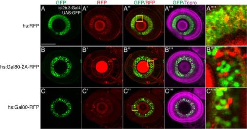
Subgroups of neurons can be distinguished by inhibiting Gal4-driven expression with a fluorescently tagged Gal80. Single slice confocal images, lateral views, anterior to left, dorsal up, of 72 hours postfertilization (hpf) eyes in Tg(isl2b.3:Gal4)zc65; Tg(UAS:GFP) embryos. Immunostaining for green fluorescent protein (GFP), green; TagRFP, red; Topro3 nuclear stain, magenta. A–A′′ ′: Embryos injected with hsp70l:TagRFP (no Gal80 expression). B–B′′ ′: Embryos injected with hsp70l:Gal80-2A-TagRFP and heat-shocked at 48 hpf show expression of TagRFP (RFP) and inhibition of Gal4-dependent expression. C–C′′ ′: Embryos injected with hsp70l:Gal80-TagRFP and heat-shocked at 48 hpf show RFP expression and concomitant inhibition of Gal4-dependent expression. (A′′ ′′–C′′ ′′) shows magnified view of the insets in (A′′–C′′), demonstrating disjoint GFP and RFP expression. Scale bar = 50 μm. |