
- Title
-
An inducible krasV12 transgenic zebrafish model for liver tumorigenesis and chemical drug screening
- Authors
- Nguyen, A.T., Emelyanov, A., Koh, C.H., Spitsbergen, J.M., Parinov, S., and Gong, Z.
- Source
- Full text @ Dis. Model. Mech.

Mifepristone-inducible liver-specific krasV12 expression in transgenic zebrafish. (A) Schematic diagram of the mifepristone-inducible LexPR system with separate liver-driver and Ras-effector constructs. The liver-specific expression of EGFP-krasV12 in double-transgenic fish (driver-effector), harboring both cassettes, is activated in trans by the LexPR activator produced from the liver-driver in the presence of mifepristone (RU486). (B–E) Representative 7-dpf fry from the liver-driver (B), Ras-effector (C) and driver-effector double transgenics (D,E). All fry were incubated with 1 µM mifepristone from 3 dpf. The upper panels show a fluorescent image and the lower panels show histology. The liver-driver fry had induced EGFP expression in the liver and a normal liver histology (B). The Ras-effector fry had mCherry expression in the lens as a transgenic marker but no induced EGFP expression in the liver, and also had a normal liver histology (C). EGFP-krasV12 expression was induced in driver-effector double-transgenic larvae at 3 dpf, causing the development of an enlarged and hyperplastic liver (D) and some individuals showed invasive EGFP-positive cells as indicated by arrows (E). Dotted areas indicate the liver regions. Scale bars: 50 μm. |

Dosage-dependent induction of krasV12 expression and liver tumor induction and regression. (A) RT-PCR analysis of transgenic krasV12 transcripts in the liver of 1-month-old driver-effector double-transgenic larvae after 3 days of mifepristone (RU486) induction at different concentrations. Induced and non-induced driver transgenics together with non-induced driver-effectors were used as negative controls. β-actin was used as loading control. 2-Off, treatment with 2 mM mifepristone followed by withdrawal for 7 or 14 days. (B) Tumor induction rates for 1-month-old driver-effector fish under different concentrations of RU486 (n=20 in each group). (C) Statistics of tumor incidence as determined by histopathological analysis on induced driver-effector transgenics after 1–4 weeks of 2 μM RU486 treatment. WT, wild type. (D) Liver tumor progression in driver-effector fish induced by 2 μM mifepristone. The four panels represent different stages of liver tumorigenesis starting from normal control liver on the left to hyperplastic liver (HL) (2 weeks), HCC (4 weeks) and hepatoblastoma in mixed HCC (8 weeks). Each panel contains brightfield and fluorescent images of gross liver morphology (top) and a relevant histological image (bottom). (E) Liver tumor regression after mifepristone withdrawal. Following treatment of 2 μM RU486 for 4 weeks, driver-effector fish developed EGFP-labeled HCC and their exposure to RU486 was then terminated. Histological sections from two representative fish after RU486 removal for 2 and 4 weeks are shown. Dotted areas showed tumor shrinkage with marked peripheral scarring, whereas asterisks indicate surrounding normal hepatic tissue. (+) and (–) indicate RU486-treated and untreated fish, respectively. Boxed areas are shown enlarged on the right. Scale bars: 100 μm. PHENOTYPE:
|

Roles of the Raf-MEK-ERK and PI3K-AKT-mTOR pathways during krasV12 tumor progression and regression. (A) Western blots using total liver proteins from induced driver-effector fish with HL or HCC, as well as regressed liver samples, to detect total Kras (F234), mutant Kras-2B (C19) isoform, phospho-ERK1/2 (P-ERK1/2), P-AKT and P-S6 proteins. The non-induced driver, induced-driver and non-induced driver-effector having normal liver (N) served as controls.β-actin, internal control for equal loading. (B) Immunohistochemical staining showing phospho-ERK1/2 and -AKT protein as well as apoptotic cells (TUNEL) during krasV12-dependent liver tumor progression and regression. Asterisks and dotted lines indicate the region of tumor scar. EXPRESSION / LABELING:
|

Suppression of liver tumorigenesis by inhibition of the Raf-MEK-ERK and PI3K-AKT-mTOR pathways. (A) Fluorescent images showing representative mifepristone-induced EGFP-krasV12 driver-effector larvae at 7 dpf treated with different inhibitors, as compared with the DMSO-treated driver and driver-effector transgenics as controls. Scale bar: 200 μm. (B) Comparison of the anti-HL effect of inhibitors in transgenic larvae at 7 dpf. The liver size of treated larvae was classified as ‘normal’ if it was the same as the liver in driver transgenics, ‘huge’ if it remained enlarged/hyperplasia, or ‘large’ if it was larger than normal liver, but smaller than hyperplasia after 4 days of inhibitor treatment. (C) Lysates extracted from whole transgenic larvae treated with the indicated inhibitors were immunoblotted for phospho-ERK (P-ERK), P-AKT and P-S6. β-actin, internal control for equal loading. (D) Proposed inhibitor treatments to suppress KrasV12-induced liver tumorigenesis via targeting both the Raf-MEK-ERK and PI3K-AKT-mTOR pathways. EXPRESSION / LABELING:
|
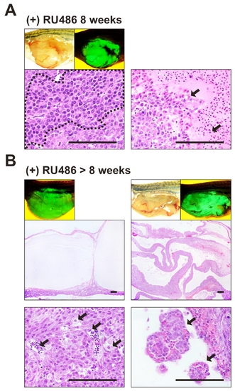
Advanced liver tumors in induced krasV12 double transgenic fish.Morphological and histological examinations of liver tumors were carried out in driver-effector fish induced with mifepristone (RU486) for 8 weeks or longer. Representative brightfiled and fluorescent images of liver morphology and histology of induced Driver-effector fish are shown. (A) One sample from 8 weeks of RU486 treatment. Histoplathological analysis revealed that hepatoblastoma (dotted area) which is made up of undifferentiated cells with large nuclei developed in mixed carcinoma in Driver-effector fish after 8 weeks of induction. In addition, these fish also showed invasion of tumor cells into blood vessels. (B) Examples from more than 8 weeks of mifepristone induction. 10% of induced driver-effector fish developed cyst fluid or malignant ascites in HCC. Detached HCC cells were found in the cysts and blood vessels. Black arrows indicated invasive tumor cells. Scale bars, 100 μm. |

Activation of both ERK and AKT pathways in advanced liver cancer of krasV12 transgenic fish. Driver-effector fish induced with mifepristone (RU486) for more than 8 weeks developed hepatoblastoma (HB) and ascites in mixed HCC. (A) Immunohistochemistry for PERK1/ 2 and P-AKT showing positive nuclear and cytoplasmic staining of both P-ERK1/2 and PAKT proteins in HB (dotted area). (B) Ascites and cyst fluid in mixed HCC also displaying strong expression of P-ERK1/2 and P-AKT protein. |