
- Title
-
Mutations in fam20b and xylt1 Reveal That Cartilage Matrix Controls Timing of Endochondral Ossification by Inhibiting Chondrocyte Maturation
- Authors
- Eames, B.F., Yan, Y.L., Swartz, M.E., Levic, D.S., Knapik, E.W., Postlethwait, J.H., and Kimmel, C.B.
- Source
- Full text @ PLoS Genet.
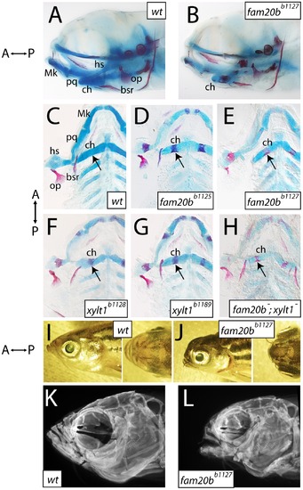
Craniofacial skeletons of mutant zebrafish larvae exhibit increased bone matrix and decreased cartilage matrix. A–H, Alcian blue/Alizarin red-stained 6 dpf larvae, entire heads viewed laterally (A,B, eyes removed) or flat-mounted, dissected pharyngeal skeletons viewed ventrally (C–H). I–L, lateral and dorsal views of entire 8-month-old heads (I,J), and corresponding lateral view images of Alizarin red fluorescence taken by optical projection tomography (OPT; K,L). Overall gross anatomy of the head and craniofacial skeleton are similar in wild types (A) and mutants (B), although staining of cartilage and bone appeared altered in mutants. Compared to wild types (C), fam20bb1125 (D), fam20bb1127 (E), xylt1b1128 (F), and xylt1b1189 (G) mutants had increased Alizarin red staining of bone (arrows) and decreased Alcian blue staining of cartilage. There was no sided-ness to the phenotype, for defective cartilage and bone appeared symmetrically on left and right sides. Double mutant fam20bb1127;xylt1b1128 larvae (H) had Alizarin red staining (arrow) similar to that seen in single mutants, but more severe loss of Alcian blue staining. Relative to wild-type siblings (I), fam20bb1127 mutant adults (J) showed foreshortened upper and lower jaws, hypoplastic midface, and bulging eyes. These overall morphological features were underlain by severe reductions to the size of the mutant craniofacial skeleton (K,L). Abbreviations: A = anterior; bsr = branchiostegal ray; ch = ceratohyal; hs = hyosymplectic; Mk = Meckel′s; op = opercle; P = posterior; pq = palatoquadrate. |
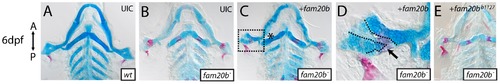
Wild-type fam20b expression rescues the fam20bb1127 mutant phenotype. A–E, Flat-mounted, dissected pharyngeal skeletons of Alcian blue/Alizarin red-stained 6 dpf larvae. Both sides of the pharyngeal skeletons are shown because the mosaic nature of the rescue experiment produced asymmetric phenotypes in injected larvae. Compared to uninjected wild-type (A) and mutant (B) controls, fam20bb1127 mutant larvae that were injected with wild-type fam20b cDNA under the control of beta-actin2 promoter (C) showed skeletal elements with rescued cartilage matrix staining by Alcian blue and decreased bone matrix staining (asterisk) by Alizarin red. D, Higher magnification of boxed region in C illustrates patches of light Alcian blue staining surrounded by heavy Alizarin red staining (arrow, dashed lines) adjacent to patches of dark Alcian blue staining surrounded by light Alizarin red staining. E, fam20bb1127 mutants embryos similarly injected with fam20bb1127 did not rescue the mutant skeletal phenotype. Abbreviations: UIC = uninjected control. |

fam20b and xylt1 are expressed in chondrocytes, but not in perichondrium. A,F, RT-PCR on wild-type extracts at ages indicated; B,C,G,H,K, whole-mount and D,E,I,J,L section in situ hybridization on wild-type embryos. RT-PCR demonstrated transcripts for fam20b (A) and xylt1 (F) from 3 hpf through 54 hpf. Frontal and lateral views of embryos stained by whole-mount in situ hybridization revealed transcripts for fam20b (B,C) and xylt1 (G,H) in developing cartilage elements of the craniofacial and pectoral fin skeletons at 55 hpf. In addition, there was diffuse expression of fam20b in the brain and specific xylt1 expression in the forebrain (fb). Horizontal section in situ hybridization localized transcripts for fam20b and xylt1 to developing chondrocytes (c) of the ceratohyal at 63 hpf (D,I) and 72 hpf (E,J), but no expression in perichondrium (pc) was detected. Lateral view of whole-mount (K) and horizontal section (L) in situ hybridization revealed expression of xylt1 in osteoblasts of the opercle (op) at 72 hpf. Abbreviations: -ctl = negative control; c = chondrocytes; ch = ceratohyal; e = eye; et = ethmoid; fb = forebrain; hpf = hours post-fertilization; hs = hyosymplectic; Mk = Meckel′s; op = opercle; pc = perichondrium; pf = pectoral fin; pq = palatoquadrate. EXPRESSION / LABELING:
|
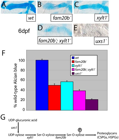
Reduction of Alcian blue staining in fam20b and xylt1 mutants is less severe than in uxs1 mutants. A–E, Alcian blue-stained 6 dpf ceratohyals. Compared to wild types (A), Alcian blue staining appeared lower in fam20bb1127 (B) and xylt1b1128 (C) mutants. Staining in fam20bb1127;xylt1b1128 double mutants (D) was decreased in comparison to single mutants, although uxs1 mutants (E) showed the greatest decrease in Alcian blue staining. F, Quantitation of Alcian blue in lysates of 6 dpf larvae revealed statistically significant changes (ANOVA p<0.0001) for each mutant class. Compared to wild-type Alcian blue staining, fam20bb1127 mutants had 50±3.0%, xylt1b1128 mutants had 57±2.0%, fam20bb1127;xylt1b1128 double mutants had 39±2.6%, and uxs1 mutants had 22±1.5%. G, Simplified pathway of PG synthesis, demonstrating that uxs1 is upstream of both xylt1, which adds xylose to a serine (Ser) residue of the core protein, and also fam20b, which phosphorylates (P) xylose in the nascent GAG chain. Abbreviations: CSPGs = chondroitin sulfate proteoglycans; HSPGs = heparan sulfate proteoglycans. PHENOTYPE:
|
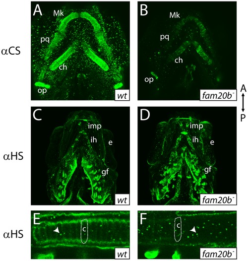
fam20b mutants display reduced proteoglycan immunoreactivity A–F, Ventral views of confocal images of embryonic heads immunostained for chondroitin sulfate proteoglycan (CSPG) at 3 dpf (A,B) or heparan sulfate PG (HSPG) at 4 dpf (C–F). Compared to abundant CSPGs detected in wild-type skeletal elements (A), fam20bb1127 mutants showed decreased CSPG immunoreactivity (B). HSPGs were abundant in gill filaments and ventral muscle groups of both wild-type (C) and fam20bb1127 mutant zebrafish (D). HSPG immunoreactivity was reduced greatly around developing chondrocytes of the fam20bb1127 mutant ceratohyal (outline in F), compared to those of wild-type siblings (outline in E). Also, the number of intracellular HSPG aggregates (arrowhead) was increased in fam20bb1127 mutant chondrocytes. Abbreviations: A = anterior; αCS = anti-chondroitin sulfate; αHS = anti-heparan sulfate; c = chondrocyte; ch = ceratohyal; e = eye; gf = gill filament; ih = interhyoideus; imp = intermandibularis posterior; Mk = Meckel′s; op = opercle; P = posterior; pq = palatoquadrate. EXPRESSION / LABELING:
PHENOTYPE:
|
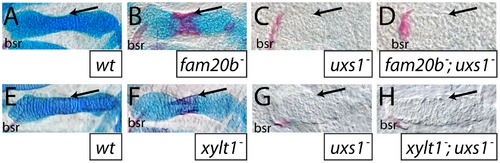
The uxs1 mutation is epistatic to cartilage and bone phenotypes of fam20b and xylt1 mutants. A–H, Alcian blue/Alizarin red-stained 6 dpf ceratohyals. Compared to wild types (A,E), cartilage matrix of fam20bb1127 (B) and xylt1b1128 (F) mutants stained less with Alcian blue, but the perichondria of fam20b and xylt1 mutants stained more with Alizarin red (arrows) than in wild types. Similar to uxs1 mutants (C,G), uxs1;fam20bb1127 (D) and uxs1;xylt1b1128 (H) double mutants showed a greater decrease in Alcian blue staining of cartilage matrix than the decrease seen in fam20b and xylt1 mutants; also, Alizarin red staining (arrows) in uxs1 single mutant and uxs1;fam20b and uxs1;xylt1 double mutant perichondria was at wild-type levels. Abbreviations: bsr = branchiostegal ray. |

fam20b and xylt1 mutants have increased perichondral bone. At 6 dpf, Alizarin red staining (see Figure 1C–1G) was visible more often in skeletal elements of fam20bb1127 (A) and xylt1b1128 (B) mutants than in wild types. Statistically significant (*, p<0.05) increases, however, were observed in more chondral bones (pq, hm, ch) than dermal bones (ept, mx). Quantitative analyses of the sum of bone areas in left and right ceratohyals (ch(l) and ch(r), dashed outlines) in whole-mount, ventral images of live Alizarin red fluorescence (C–E) revealed statistically significant (*, p<0.05) increases in fam20bb1127 (F) and xylt1b1128 (G) mutants, compared to wild-type siblings at 6 dpf. Abbreviations: A = anterior; ch = ceratohyal; ept = entopterygoid; hm = hyomandibular; l = left; mx = maxilla; pq = palatoquadrate; P = posterior; r = right. PHENOTYPE:
|
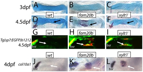
Precocious bone formation and osteoblast differentiation in perichondria of fam20b and xylt1 mutants. A–F, Alcian blue/Alizarin red-stained ceratohyals. G–I, live Alizarin red fluorescence of fam20bb1127;Tg(sp7:EGFP)b1212 and xylt1b1128;Tg(sp7:EGFP)b1212 larvae. J–O, whole-mount in situ hybridization of 4 dpf ceratohyals. No signs of Alizarin red staining were observed at 3 dpf in ceratohyals of wild types (A), or fam20bb1127 (B) or xylt1b1128 (C) mutants. By 4.5 dpf, Alizarin red staining was still absent from ceratohyals of wild-type larvae (D, arrow), although it was detected in ceratohyals of fam20bb1127 (E, arrow) and xylt1b1128 (F, arrow) mutants. Both GFP expression and Alizarin red staining were at background levels in the perichondrium of the ceratohyal in 4.5 dpf wild types (G, arrow), but were obvious in the fam20bb1127 and xylt1b1128 mutant perichondria (H,I, arrow). At 4 dpf, col10a1 expression was not detected in the ceratohyal perichondrium of wild types (J, arrow), but was expressed highly in fam20bb1127 (K, arrow) and xylt1b1128 (L, arrow) mutant perichondria. Abbreviations: bsr = branchiostegal ray. EXPRESSION / LABELING:
PHENOTYPE:
|

Early molecular markers of chondrocyte maturation in fam20b and xylt1 mutants. A–R, horizontal section in situ hybridization of developing ceratohyals. The levels of runx2b transcripts appeared higher in chondrocytes (c) and perichondrium (pc) of the ceratohyal in fam20bb1127 (B) and xylt1b1128 (C) mutants, than in wild types (A) at 72 hpf. Expression of col10a1 was abundant in chondrocytes (c) and perichondrium (pc) of the ceratohyal in fam20bb1127 (E) and xylt1b1128 (F) mutants, but was not detectable in wild types (D) at 83 hpf. Levels of ihha and ihhb transcripts were up-regulated in chondrocytes (c) of the ceratohyal in fam20bb1127 (H,K) and xylt1b1128 (I,L) mutants, but were not detectable in wild types (G,J) at 72 hpf. Transcript levels for ptch2 were increased in the perichondrium of fam20bb1127 (N) and xylt1b1128 (O) mutants, compared to wild types (M) at 72 hpf, whereas no obvious differences in levels of ptch1 expression in perichondria were apparent (P–R). Abbreviations: c = chondrocytes; pc = perichondrium. EXPRESSION / LABELING:
PHENOTYPE:
|

Ultrastructural evidence of premature chondrocyte hypertrophy and aberrant matrix production in xylt1 mutants. A–D, transmission electron micrographs of 84 hpf ceratohyals. At 5600X magnification, chondrocytes in the central region of the wild-type ceratohyal displayed abundant rough ER, Golgi, and mitochondria in the cytoplasm (A). Similar views of xylt1 mutant chondrocytes at the same age and comparable positions within the developing ceratohyal showed cytoplasmic clearing and tremendous reduction in biosynthetic organelles (B). Mutant chondrocytes also appeared to be separated by less extracellular matrix. While wild-type (C) and mutant (D) extracellular matrix demonstrated fibrillar collagens at 31000X magnification, mutant matrix showed tighter fibril packing and also contained more amorphous electron-dense structures (*) in pericellular matrix than seen in wild types. Abbreviations: cyt = cytoplasm; ecm = extracellular matrix; nuc = nucleus. Scale bars: A,B = 2 μm; C,D = 500 nm. PHENOTYPE:
|
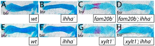
The ihha mutation is epistatic to bone, but not cartilage, phenotypes of fam20b and xylt1 mutants. A–H, Alcian blue/Alizarin red-stained 6 dpf ceratohyals. Alcian blue staining was comparable between wild-type (A,E) and ihha mutant (B,F) cartilages, but was decreased in fam20bb1127 (C) and xylt1b1128 (G) single mutant, and in fam20bb1127;ihha (D) and xylt1b1128;ihha (H) double mutant, cartilages. Alizarin red staining of fam20bb1127 (C) and xylt1b1128 (D) chondral bones was abundant, while no such staining was observed in wild types (A,E), ihha mutants (B,F), or fam20bb1127;ihha (D) and xylt1b1128;ihha double mutants (H). Abbreviations: bsr = branchiostegal ray. |

Exogenous wild-type fam20b expression does not alter skeletal phenotypes of wild types or xylt1b1128 mutants, but can rescue cartilage of fam20bb1125 mutants with a single induction at 55 hpf. A–E, dissected, flat-mounted pharyngeal skeletons of Alcian blue/Alizarin red-stained (A,B) and Alcian blue-stained (C–E) larvae. Injection of larvae with wild-type fam20b under control of the beta-actin2 promoter did not alter skeletal phenotypes of wild types (A) or xylt1b1128 mutants (B). Injection of larvae with wild-type fam20b under control of the hsp70l promoter rescued cartilage matrix production in fam20bb1125 mutants when heat shocked at 55 hpf (E), compared to injected fam20bb1125 embryos that did not undergo heat shock (D), while heat shock had no effect on uninjected control larvae (C). Abbreviations: HS = heat shock; UIC = uninjected control. |

Cartilage proteoglycan secretion initiates at the same time in xylt1 mutants and wild types. Alcian blue matrix was not apparent in pharyngeal arch 1 or 2 of wild types (A) and xylt1b1128 mutants (B) at 48 hpf. Staining in the trabeculae of the neurocranium is evident in both wild types and xylt1b1128 mutants. Alcian blue staining was obvious in pharyngeal arches of both wild types (C) and xylt1b1128 mutants (D) at 60 hpf. Abbreviations: ch = ceratohyal; pa1 = pharyngeal arch1; pa2 = pharyngeal arch 2; pq = palatoquadrate; t = trabeculae. |