
- Title
-
liver-enriched gene 1a and 1b Encode Novel Secretory Proteins Essential for Normal Liver Development in Zebrafish
- Authors
- Chang, C., Hu, M., Zhu, Z., Lo, L.J., Chen, J., and Peng, J.
- Source
- Full text @ PLoS One
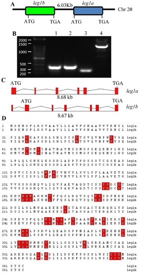
Leg1a and Leg1b are two closely related homologs in zebrafish. (A) Diagram showing the arrangement of leg1a and leg1b on chromosome 20. (B) RT-PCR showing the expression of leg1a and leg1b in adult liver. Primer pairs amplifying leg1a are derived from exon1 of leg1a (lanes 1 and 2), leg1b are from exon1 and exon2 of leg1b (lanes 3 and 4). Lanes 1 and 3: cDNA as template; lanes 2 and 4: genomics DNA as template. (C) Diagram showing leg1a and leg1b genomic structures. Red box: exon; uneven line: introns. (D) Alignment of Leg1a and Leg1b amino acid sequences to highlight the 39 different amino acids between them (boxed in red). |
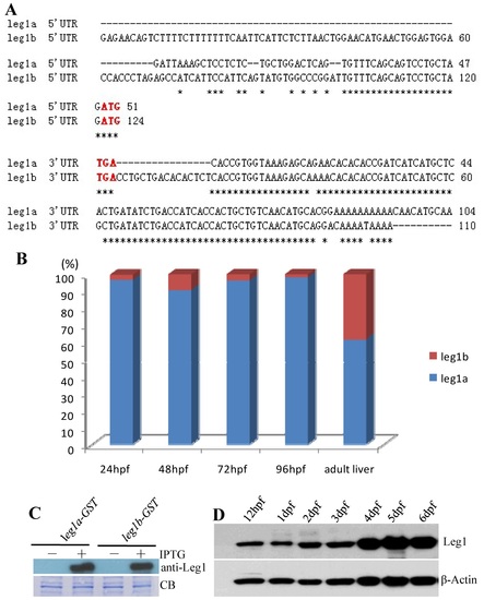
leg1a and leg1b are differentially expressed. (A) Alignment of 5′- and 3′-UTRs of leg1a and leg1b transcripts, respectively. Asterisk highlights identical bases. Translation start codon ATG and stop codon TGA are lettered in red. (B) Comparison of leg1a and leg1b expression at different developmental stages as indicated. Data are presented in percentage (blue bar: leg1a; red bar: leg1b). (C) Anti-Leg1 monoclonal antibody recognizes both Leg1a and Leg1b which were induced to express in E. coli by isopropyl β-D-1-thiogalactopyranoside (IPTG). (D) Western blotting analysis of total Leg1 (Leg1a+Leg1b) expression in embryos at stages as indicated. |
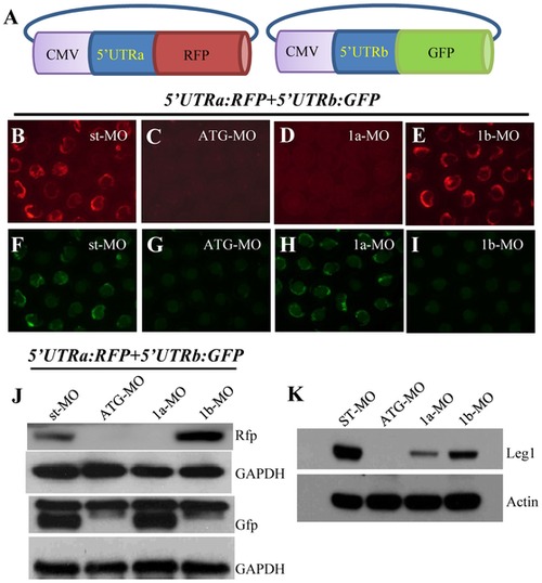
Knockdown of Leg1a or Leg1b protein expression with their specific morpholinos. (A) Diagram showing construction of leg1a-52-UTR:rfp (52UTRa:RFP) and leg1b-52-UTR:gfp (52UTRb:GFP) plasmids. (B–I) 52UTRa:RFP and 52UTRb:GFP were mixed and co-injected with st-MO (B, F), leg1-MOATG (C, G) or leg1a-MO (D, H), or leg1b-MO (E, I). Rfp (B–E) and Gfp (F–I) fluorescence was visualized under a Nikon fluorescence microscope. (J) Western blotting analysis of Rfp and Gfp proteins in the injected embryos as indicated using antibodies against Rfp and Gfp, respectively. (K) Western blotting analysis of total Leg1 in different morphants as indicated. |
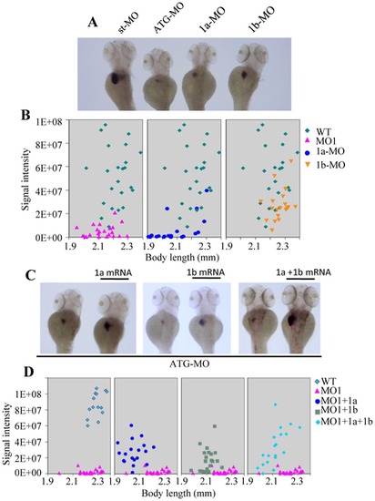
Both Leg1a and Leg1b are essential for normal liver development. (A) leg1-MOATG (ATG-MO), leg1a-MO (1a-MO) and leg1b-MO (1b-MO) all resulted in small liver phenotype but with different severity. (B) Statistical analysis of liver size in leg1-MOATG (ATG-MO), leg1a-MO (1a-MO) or leg1b-MO (1b-MO) morphants based on WISH signal intensity of fabp10a. Signal intensity (Y-axis) was plotted against body length (X-axis). 20–23 embryos were used for statistical analysis in each case. (C) WISH using fabp10a probe to examine rescue of the small liver phenotype caused by leg1-MOATG with leg1a (1a mRNA), leg1b (1b mRNA) mRNA, or combination of leg1a and leg1b mRNA (1a+1b mRNA). (D) Statistical analysis of liver size in each individual leg1-MOATG morphant based on WISH signal intensity of fabp10a. 19–23 embryos were used for statistical analysis in each case except for WT (14 embryos were used). PHENOTYPE:
|
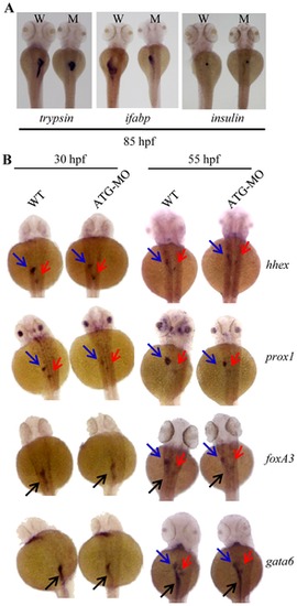
Depletion of Leg1 affects the expansion but not initiation of the liver, exocrine pancreas and intestinal tub. (A) WISH using trypsin, ifabp and insulin as probes to examine the development of exocrine pancreas (trypsin), intestine (ifabp) and endocrine pancreas (insulin) in WT (W) and the leg1-MOATG morphant (M) embryos at 85 hpf. (B) WISH using early hepatic markers hhex and prox1 and early endoderm markers foxa3 and gata6 on WT and leg1-MOATG morphant (ATG-MO) embryos at 30 hps and 55 hpf, respectively. Blue arrow: liver; red arrow: pancreas; black arrow: intestine. PHENOTYPE:
|
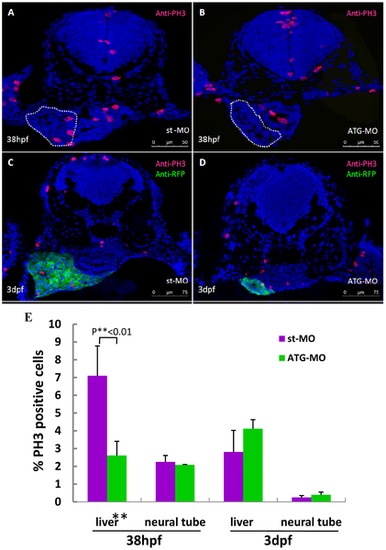
Hypoplastic liver in leg1-MOATG morphants is caused by cell cycle arrest during the liver budding stage. (A, B) Immunostaining using an anti-PH3 antibody on st-MO (A) and ATG-MO (B) morphants at 38hpf. Liver primordium is circled with a white dotted line. (C, D) Immunostaining using antibodies respectively against PH3 (red) and RFP (green) on cross-sections from st-MO (C) and ATG-MO (D) morphants in the Tg(lfabp: DsRed; elaA: EGFP) background at 3dpf. (E) Statistical analysis revealed that the ratio of PH3-positive cells was significantly reduced in the liver primordium of ATG-MO morphants at 38 hpf, but no significant difference in the ratio of PH3-positive cells was observed in the liver of ATG-MO morphants at 3dpf. Data was obtained by counting PH3-positive cells versus total cells in a specific organ (e.g. liver) in sections from at least 3 st-MO embryos and 3 ATG-MO morphants at each stage, respectively (Table S1). |