
- Title
-
An apo-14 promoter-driven transgenic zebrafish that marks liver organogenesis
- Authors
- Wang, R., Li, Z., Wang, Y., and Gui, J.F.
- Source
- Full text @ PLoS One
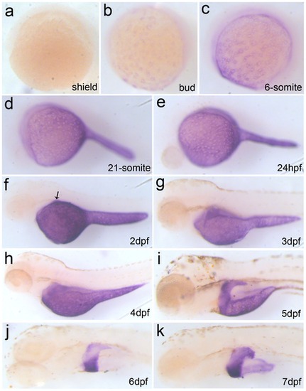
Expression pattern of endogenous Apo-14 during zebrafish embryogenesis by whole-mount in situ hybridization. (a) shield (6 hpf); (b) bud (10 hpf); (c) 6-somite (12 hpf); (d) 21-somite(20 hpf); (e) 24 hpf; (f)2 dpf larva; (g) 3 dph larva; (h) 4 dph larva; (i) 5 dph larva; (j) 6 dph larva; (k) 7 dph larva. (a) dorsal to the right; (b–k) anterior to the left. The arrow indicates positive signals for the liver primordium. |
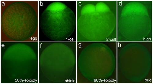
Observation of maternal expression and post-fertilization translocation of Apo-14 promoter-driven GFP in transgenic zebrafish line Tg(Apo14: GFP). (a) unfertilized egg; (b) 1-cell embryo; (c) 2-cell embryo; (d) high blastula embryo; (e) 50%-epiboly embryo; (f) shield embryo; (g) 90%-epiboly embryo; (h) bud stage embryo. |
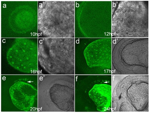
Onset expression and developmental behavior of Apo-14 promoter-driven GFP in early heterozygous embryos produced by out-crossing the Tg(Apo14: GFP) male to the wild type female. (a, a′) bud embryo at 10 hpf; (b, b′) 5-somite embryo at 12 hpf; (c, c′) 14-somite embryo at 16 hpf; (d, d′) 17 hpf embryo; (e, e′) 20 hpf embryo; (f, f′) 24 hpf embryo. Arrows point to the liver primordium. The images (a–f) show the green fluorescence, and (a′-f′) the corresponding bright images. |
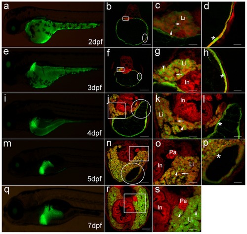
Dynamic GFP expression pattern in liver morphogenesis of Tg(Apo14: GFP) from 2dpf to 7dpf. (a, e, i, m, q) show the left lateral view of Tg(Apo14: GFP) larvae from 2dpf to 7dpf. (b, f, j, n, r) show the cross sections through intestine bulb and liver of the larvae at same stage as the left columns. Scale bar: 150 μm. (c, g, k, o, s) are magnifications of the corresponding frame areas in (b, f, j, n, r), which highlight the GFP expression in the hepatocytes. Scale bar: 30 μm. (d, h, l, p) are magnifications of the corresponding circle areas in (b, f, j, n), which highlight the GFP expression on the YSL. Scale bar: 30 μm. Green fluorescence is emitted from the Apo-14 promoter-driven GFP, and red fluorescence is stained by PI for showing the nuclear position. Arrowheads point to hepatocytes, arrows mark sinusoids, and the asterisks delineate the GFP-expressed nuclei on YSL. Li: liver; In: intestine bulb; Pa: pancreas. |
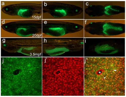
Outgrowth of the third lobe of liver from 15dpf to 20dpf and liver sections of adult fish of Tg(Apo14: GFP). (a-c) 15 dpf larvae; (d-f) 20 dpf larvae; (g-i) 3.5 mpf adult fishes. (a, d, g) are the left lateral views showing left lobe of the liver. (b, e, h) are the right lateral views showing right lobe of the liver. (c, f, i) are the central views showing ventral lobe of the liver. Anterior is to the left. (j, j′, j′′) are the cross sections through the liver of 3.5 mpf adult fish. (j) shows the green GFP fluorescence in the hepatocytes, (j′) shows the red fluorescence stained by PI, (j′′) is the mergence of the green and red fluorescence. Arrowhead points to the hepatocyte, arrow marks sinusoid, and the asterisk delineates the central vien. Scale bar: 30 μm. |