
- Title
-
Sublethal exposure to crude oil during embryonic development alters cardiac morphology and reduces aerobic capacity in adult fish
- Authors
- Hicken, C.E., Linbo, T.L., Baldwin, D.H., Willis, M.L., Myers, M.S., Holland, L., Larsen, M., Stekoll, M.S., Rice, S.D., Collier, T.K., Scholz, N.L., and Incardona, J.P.
- Source
- Full text @ Proc. Natl. Acad. Sci. USA
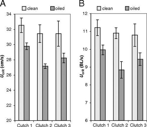
Reduced swimming performance in adult fish exposed to oiled gravel effluent as embryos. Ucrit was measured as described in Materials and Methods. Mean Ucrit (±SE) for fish embryonically exposed to effluent from clean gravel (light gray bars, n = 5) or oiled gravel (dark gray bars, n = 9) is given as an absolute speed (A; cm/s) or relative to body length (B; BL/s). Statistical analysis is discussed in the text. PHENOTYPE:
|

Embryonic oil exposure changes the cross-sectional dimensions of the adult cardiac ventricle. (A) Representative transverse section of an adult zebrafish heart showing the two main chambers (atrium and ventricle) and the bulbus arteriosus (ba). Arrows indicate locations of length and width measurements. (B) Box plots with individual data points showing length/width ratios for fish embryonically exposed to clean gravel effluent (n = 16) or oiled gravel effluent (n = 17). (Scale bar, 0.5 mm.) |

CYP1A immunofluorescence in embryos exposed to gravel effluents. Embryos were exposed from 4 to 48 hpf in either clean (A) or oiled-gravel effluent (B–F), fixed and processed for whole-mount CYP1A (green) and myosin heavy chain (red) immunofluorescence as described under Materials and Methods. The myosin heavy chain antibody recognizes both skeletal and cardiac muscle and serves as an internal control for permeabilization. Images in A and B are epifluorescent, C and D are confocal stack projections, and E and F are confocal optical sections. Embryos from clutch 2 exposed to clean (A) or oiled (B) gravel effluent. (C and D) CYP1A immunofluorescence in the epidermal cells on the head of embryos from clutch 2 and clutch 3, respectively. Eye and pectoral fins (pf) are indicated. (E and F) CYP1A immunofluorescence in endocardial cells (arrowheads) of the cardiac atrium (a) and ventricle (v), and epithelium (arrows) over the pericardial region. (Scale bars: 100 μm.) |

Morpholino knockdown of cyp1a enhances PAH cardiotoxicity, increasing the severity of pericardial edema. Representative images of embryos uninjected and exposed to oiled gravel effluent with 20 ppb ΣPAH (A), injected with standard control morpholino and exposed to 20 ppb ΣPAH (B), injected with cyp1a morpholino and exposed to 20 ppb ΣPAH (C), and injected with cyp1a morpholino and exposed to 9 ppb ΣPAH (D). (Scale bar: 200 μm.) PHENOTYPE:
|