
- Title
-
The deubiquitylating enzyme Cops6 regulates different developmental processes during early zebrafish embryogenesis
- Authors
- Tse, W.K., You, M.S., Ho, S.H., and Jiang, Y.J.
- Source
- Full text @ Int. J. Dev. Biol.

Zebrafish cops6 expression at early developmental stages. cops6 started its expression at the 1-cell stage and was ubiquitously expressed throughout early developmental stages. (A) RT-PCR results. (B-F) In situ hybridization results of embryos at different developmental stages: (B) 1-cell; (C) 4-cell; (D) 80% epiboly; (E) 12-somite and (F) prim-5. Throughout early development, cops6 mRNA was expressed at similar levels and no specific expression pattern was observed. Scale bar: 150 μm (B-F). |
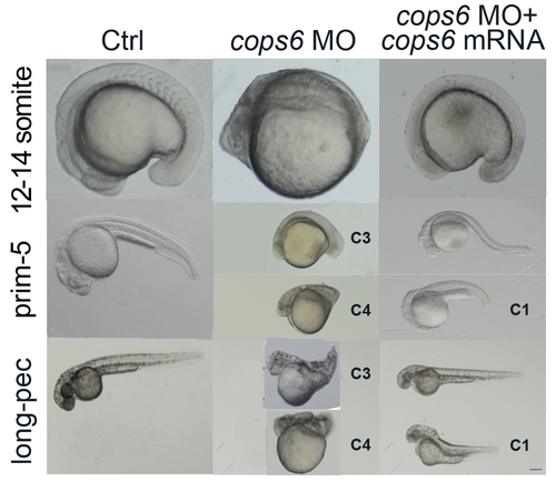
Phenotypes of cops6 morphants. cops6 morphants in an AB wild-type background showed dorsalized phenotypes at different stages: (B) 12-14 somite, (E) prim-5 and (H) long-pec. They all showed the reduced development of the posterior part, which is a feature of dorsalization. C3 and C4 dorsalized phenotypes are shown in (E,H). The dorsalized phenotype caused by cops6 knockdown was rescued by coinjection of cops6 mRNA with MO (C,F,I). More than 80% of dorsalized embryos were rescued back to a phenotype similar to wild-type, while the remaining ones showed a much milder phenotype, C1, in (F,I). Scale bar: 100 μm (A,B), 120 μm (C), 80 μm (D), 150 μm (E,F,I), 90 μm (G) and 135 μm (H). PHENOTYPE:
|

Zebrafish cops6 is required for dorsoventral patterning and convergent extension movement in early development. cops6 morphants showed a wider expression pattern of chd, marked with arrows (A,B), but a narrower expression pattern of eve1, marked with arrows (C,D); animal pole views, dorsal towards the right at 60-75% epiboly stage. The dlx3 expression domain in (F) cops6 morphants was expanded at the 1-4 somite stage when compared to (E) control. Somite morphology of (G) wild-type and (H) cops6 morphants at the 12-14 somite stage. Lateral expansion of somite muscles was apparent (green dot lines). Furthermore, myoD expression in (J) cops6 morphants was widened and severely disrupted (red asterisk) when compared to (I) control. pax2a expression at the 12-14 somite stage (K,L) lateral view and (K–L, insert) dorsal view. Blue dotted lines represent the distance between the mid-hindbrain boundary and otic vesicles in the lateral view; while red dotted lines represent this distance in the dorsal view. Shortening of the lateral distance was found in cops6 morphants. On the other hand, white dotted lines indicated the distance between the two otic vesicles, where lengthening of the ventral distance was found in cops6 morphants. mhb: mid-hindbrain boundary; ot: otic vesicle. In all photos, the head is to the left. Scale bar: 70 μm (A-D and G-J), 25 μm (E,F) and 150 μm (K,L). EXPRESSION / LABELING:
PHENOTYPE:
|
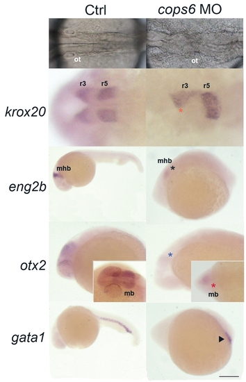
Zebrafish cops6 plays a role in brain development, but not in blood vessel formation. (A,B) cops6 morphants in an AB wild-type background showed a brain defect at the prim-5 stage. The hindbrain was not well formed in (B) cops6 morphants when compared to (A) control; dorsal view. (C,D) Expression of krox20, a marker of rhombomeres three and five; dorsal view. Reduced size in rhombomere three (orange asterisk) and fused rhombomere five expression due to unfolded hindbrain were found. (E,F) eng2b expression that indicates the mid-hindbrain boundary (mhb) was reduced in cops6 morphants (black asterisk). (F) cops6 morphants had a smaller mhb when compared to (E) control; lateral view. (G,H) otx2 expression was reduced in cops6 morphants (blue asterisk in lateral view; red asterisk in dorsal view). The size of the midbrain was decreased in (H) cops6 morphants in comparison to (G) control. (I,J) Presumptive blood marker (gata1) did not show any significant differences between (I) control and (J) cops6 morphants. They both form a normal blood island (triangle mark), which suggests that cops6 is not required for blood development. mb: midbrain; mhb: mid-hindbrain boundary; ot: otic vesicle; r3/r5: rhombomere three/five. All photos are with head to the left. Scale bar: 48 μm (A,B), 100 μm (C,D), 265 μm (E,I), 150 μm (F,J) and 170 μm (G,H). |
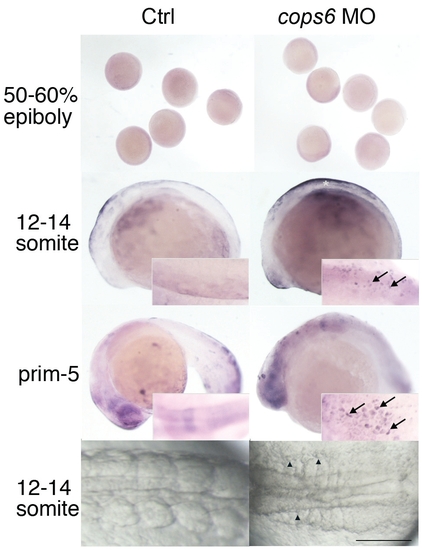
Zebrafish cops6 plays an anti-apoptotic role in early development. TUNEL assay of wild-type embryos and cops6 morphants at different developmental stages. There were no significant differences between (A) control and (B) cops6 morphants at 50-60% epiboly stage. At the 12-14 somite stage, (D) cosp6 morphants showed increased positive staining of apoptotic cells in the trunk region (white asterisk) when compared to (C) control; lateral view. Apoptotic cells (arrows) at the same stage, dorsal view (insert), were found in (D) cops6 morphants, but not in (C) control. Afterwards, at (E,F) prim-5 stage, increased numbers of apoptosis-positive stained cells (arrows) were found only in (F) cops6 morphants. (G,H) At the 12-14 somite stage, the apoptotic dying cells, exhibiting a rounded-up cell body (arrowheads), were found in (H) cops6 morphants. Scale bar: 1250 μm (A,B), 210 μm (C-F) and 150 μm (G,H). PHENOTYPE:
|