
- Title
-
Pax2/8 proteins coordinate sequential induction of otic and epibranchial placodes through differential regulation of foxi1, sox3 and fgf24
- Authors
- Padanad, M.S., and Riley, B.B.
- Source
- Full text @ Dev. Biol.
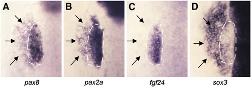
Spatial domains of otic and epibranchial markers at 12 hpf. Dorsal views showing expression of pax8 (A), pax2a (B), fgf24 (C) and sox3 (D) in wild-type embryos at 12 hpf. Otic domains (white dashed lines) and epibranchial domains (black arrows) are indicated. Unlike the other genes, fgf24 expression is limited to the otic domain. EXPRESSION / LABELING:
|
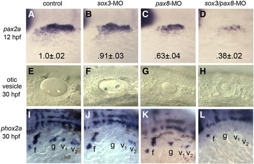
pax8 and sox3 interact in otic and epibranchial induction. (A–D) pax2a expression in the otic/epibranchial domain at 12 hpf in a control embryo (A), sox3 morphant (B), pax8 morphant (C) and sox3–pax8 double morphant (D). Numbers indicate normalized values for the mean ± standard deviation of the area of the pax2a expression domain (n = 10 specimens for each background). Area was calculated by outlining otic–epibranchial domains in Photoshop and measuring the number of pixels within. Differences between the morphants and the control were highly significant (p<.0005) as determined by t-tests. (E–H) Otic vesicles at 30 hpf in a live control embryo (E), sox3 morphant (F), pax8 morphant (G) and sox3–pax8 double morphant (H). (I–L) Expression of phox2a at 30 hpf in a control embryo (I), sox3 morphant (J), pax8 morphant (K) and sox3–pax8 double morphant (L). Positions of the facial ganglion (f) glossopharyngeal ganglion (g), and vagal ganglia (v1 and v2) are indicated. All images show lateral views with anterior to the left. EXPRESSION / LABELING:
PHENOTYPE:
|
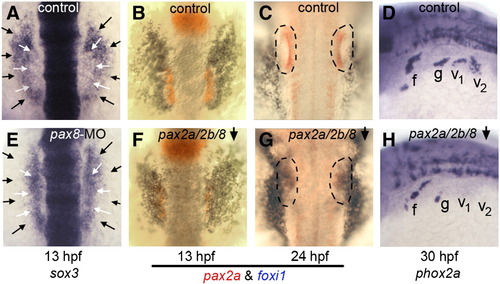
Requirement for pax2/8 in otic and epibranchial development. (A, E) Expression of sox3 at 13 hpf in a control embryo (A) and pax8 morphant (E). White arrows indicate the lateral edges of the otic domain and black arrows indicate the edges of the epibranchial domain. (B, C, F, G) Two color in situ hybridization of embryos at 13 hpf (B, F) and 24 hpf (C, G) showing expression of pax2a (red) and foxi1 (blue). Outlines indicate the otic vesicle (C) or vestigial otic region (G). Expression patterns are shown in control embryos (B, C) and pax2a/pax2b/pax8-deficient embryos (F, G). (D, H) Expression of phox2a at 30 hpf in a control embryo (D) and pax2a/2b/8-deficient embryo (H). Images show dorsal views with anterior to the top (A–C, E–G); dorsolateral views with anterior to the left and dorsal to the top (D, H). Positions of the facial ganglion (f), glossopharyngeal ganglion (g) and vagal ganglia (v1 and v2) are indicated. EXPRESSION / LABELING:
PHENOTYPE:
|
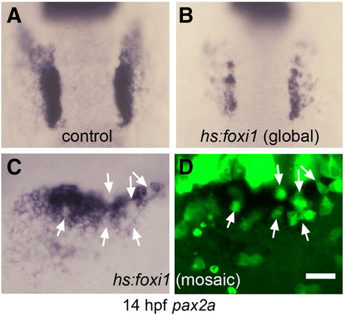
Misexpression of foxi1 inhibits otic development. (A, B) Expression of pax2a at 14 hpf in a control embryo (A) and a hs:foxi1 transgenic embryo (B) heat shocked at 11 hpf. (C, D) Expression of pax2a at 14 hpf in a mosaic embryo as seen under bright field (C) and fluorescence imaging (D). The mosaic was produced by transplanting lineage-labeled hs:foxi1 transgenic cells (green fluorescence) into a non-transgenic host. The embryo was heat shocked at 11 hpf to activate the transgene. Note the absence of pax2a expression in transgenic cells (white arrows). Images show dorsal views with anterior to the top (A–B); lateral views with anterior to the left (C–D). Scale bar, 50 μm (A, B), 25 μm (C, D). EXPRESSION / LABELING:
PHENOTYPE:
|
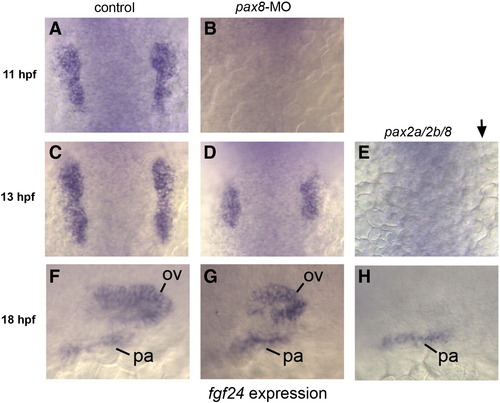
pax2/8 regulates otic expression of fgf24. (A, C, F) fgf24 expression in the otic placode in control embryos at 11 hpf (A), 13 hpf (C) and 18 hpf (F). (B, D, G) fgf24 expression in pax8 morphants at 11hpf (B), 13 hpf (D) and 18 hpf (G). Expression of fgf24 is lost from preotic placodes in pax8 morphants at 11 hpf (B) and is reduced in pax8 morphants at 13 hpf (D) and 18 hpf (G). (E, H) noi (pax2a) mutants co-injected with pax8-MO and pax2b-MO showing loss of otic expression of fgf24 at all time points. Expression in pharyngeal (pa) arches and the otic vesicle (ov) is indicated. Images show dorsal views with anterior to the top (A–E); lateral views with anterior to the left and dorsal to the top (F–H). EXPRESSION / LABELING:
|
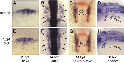
fgf24 is required for epibranchial development. (A, E) Expression of pax8 at 11 hpf in a control embryo (A) and fgf24 morphant (E). (B, F) Expression of sox3 at 14 hpf in a control embryo (B) and fgf24 morphant (F). White arrows indicate the lateral edges of the otic domain and black arrows indicate the lateral edges of the epibranchial domain. (C, G) Two color in situ hybridization showing pax2a (red) and foxi1 (blue) in a control embryo (C) and fgf24 morphant (G) at 14 hpf. Positions of otic placodes (op) are indicated. (D, H) Expression of phox2a at 30 hpf in a control embryo (D) and fgf24 morphant (H). Positions of the facial ganglion (f), glossopharyngeal ganglion (g) and vagal ganglia (v1 and v2) are indicated. Images show dorsolateral views with anterior to the left (A, D, E, H); dorsal views with anterior to the left and dorsal to the top (B, C, F, G). EXPRESSION / LABELING:
PHENOTYPE:
|
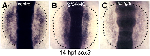
Response of sox3 to elevated Fgf signaling. Expression of sox3 at 14 hpf in a control embryo (A), fgf24 morphant (B) and hs:fgf8 transgenic embryo (C). The lateral edges of the prospective epibranchial domain are indicated (dashed lines). All embryos were heat shocked at 11 hpf. EXPRESSION / LABELING:
PHENOTYPE:
|
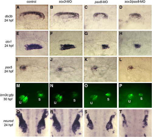
Patterning of the otic vesicle in sox3–pax8 morphants. Expression of dlx3b, otx1, pax5, and neurod in the otic vesicle of control embryos (A, E, I, Q), sox3 morphants (B, F, J, R), pax8 morphants (C, G, K, S) and sox3–pax8 double morphants (D, H, L, T) at 24 hpf. (M–P) brn3c:GFP expression at 30 hpf in a control embryo (M), sox3 morphant (N), pax8 morphant (O) and sox3–pax8 double morphant (P). Positions of utricular (u) and saccular (s) maculae are indicated. Images show dorsolateral views with anterior to the left (A–P); dorsal views with anterior to the top (Q–T). |
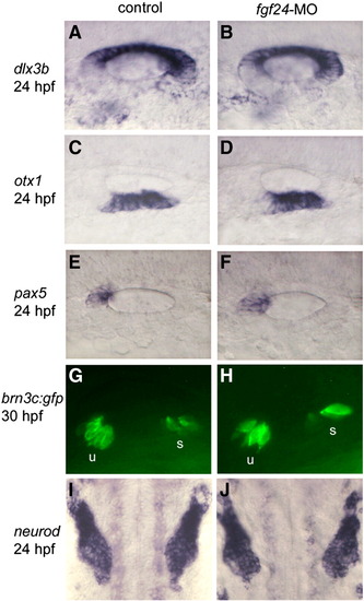
Patterning of the otic vesicle in fgf24 morphants. Expression of dlx3b, otx1, pax5, and neurod in the otic vesicle of control embryos (A, C, E, I) and fgf24 morphant (B, D, F, J) at 24 hpf. brn3c:GFP expression at 30 hpf in control embryo (G) and fgf24 morphant (H). Positions of utricular (u) and saccular (s) maculae are indicated. Images show dorsolateral views with anterior to the left (A–H); dorsal views with anterior to the top (I, J). |
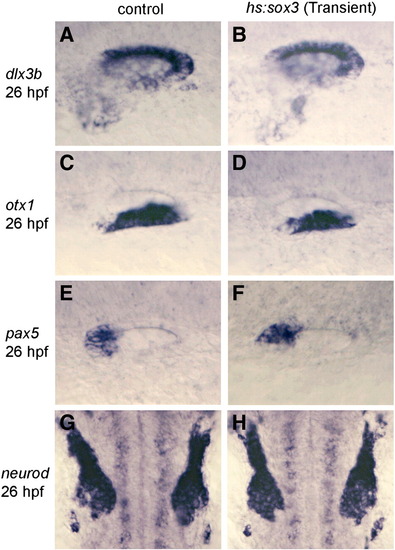
Patterning of the otic vesicle following misexpression of sox3. Expression of dlx3b, otx1, pax5, and neurod in control embryos (A, C, E, G) and in embryos transiently transfected with hs:sox3 (B, D, F, H). Embryos were heat shocked for 30 min at 39 °C beginning at 11.5 hpf, after which embryos were maintained at 33 °C. These conditions yield maximal expression of the heat shock vector for 90 min, followed by sustained lower level misexpression until fixation at 26 hpf. |
Reprinted from Developmental Biology, 351(1), Padanad, M.S., and Riley, B.B., Pax2/8 proteins coordinate sequential induction of otic and epibranchial placodes through differential regulation of foxi1, sox3 and fgf24, 90-98, Copyright (2011) with permission from Elsevier. Full text @ Dev. Biol.