
- Title
-
Wnt/PCP signaling controls intracellular position of MTOCs during gastrulation convergence and extension movements
- Authors
- Sepich, D.S., Usmani, M., Pawlicki, S., and Solnica-Krezel, L.
- Source
- Full text @ Development

Embryonic regions examined, methods for MTOC position measurement and some functions of centrin-labeled MTOCs. (A) In a midgastrulation embryo, Wnt/PCP signaling is required in cells of the dorsal midline (D) for convergence and extension, but not in lateral regions. The regions within the dotted white line were examined for MTOC angle. Scale bar: 200 μm. (B) Late gastrulation embryo. Lateral mesodermal cells flank the notochord and require Wnt/PCP signaling to migrate efficiently. (C) Membrane (red) and centrosome (green) labeling. Cells were oriented so that anterior of the embryo was oriented towards the top of the frame during measurement. Dorsal (D) and ventral (V) in the embryo are indicated. (D) Nucleus is visible in DIC optics with a green dot denoting an overlain MTOC. Dotted black line indicates orientation of MTOC relative to the nucleus. Distance between MTOC and nucleus was not measured. (E) Illustration of how MTOC position was measured relative to the nucleus N and the embryonic body axes. Angle measured is indicated by the red line. (F) Centrosomes (Xenopus centrin-Cherry, white arrowhead) in projected z-stack have a membrane connection to the cell membrane. (CAAX-EGFP, blue arrowhead). (G) Centrosomes move in cells. Paths followed for 10 minutes start at the square. (H) Short cilia (red, acetylated tubulin, white arrowhead) colocalize with some centrosomes (eGFP-Xcentrin, blue arrowheads). Scale bars: 10 μm. |

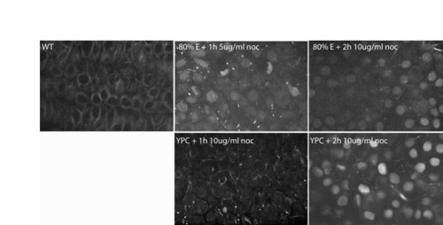
Intact microtubules are not required for Pk maintenance at anterior cell membrane, but are need for establishment of anterior localization. In all panels, embryos are one-somite stage and anterior is upwards; cells are presumptive mesoderm (at least one cell layer away from the enveloping layer cells or clearly below ectoderm/mesoderm boundary). Cells are labeled with Histone2B-RFP, Membrane-RFP and Pk-GFP. (A) Wild-type embryo grown without nocodazole, (B) grown in 10 μg/ml nocodazole for 1 hour, starting at late gastrulation or (C) grown in 10 μg/ml nocodazole for 2 hours, starting at midgastrulation. (D) The location of Pk-GFP labeling was noted for axial mesoderm, presomitic mesoderm (PSM) and ectoderm in wild-type and 1 hour nocodazole-treated embryos (n=7, each condition). Cellular localization is largely stable to 1 hour treatment. |
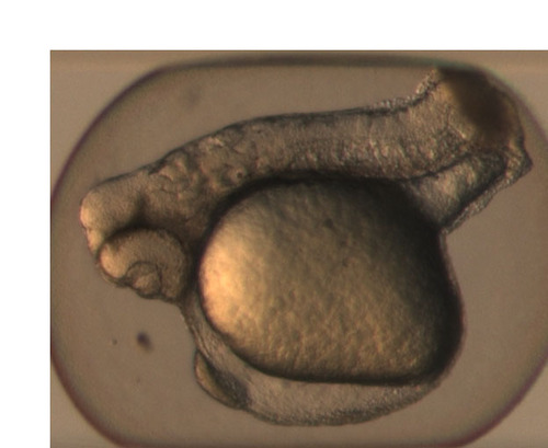
Xdd1 dose effects C&E not dorsal fate. Zebrafish embryos injected with 400 pg Xdd1 at the one-cell stage have shortened axes and cyclopia, but have normal amounts of dorsal tissues. |
Nocodazole quickly depolymerizes MT and eventually rounds cells. Embryos from transgenic line expressing a GFP-microtubule-binding protein. Confocal images were obtained at the one-somite stage. |