
- Title
-
Zebrafish numb and numblike are involved in primitive erythrocyte differentiation
- Authors
- Bresciani, E., Confalonieri, S., Cermenati, S., Cimbro, S., Foglia, E., Beltrame, M., Di Fiore, P.P., and Cotelli, F.
- Source
- Full text @ PLoS One

The morpholino can block the translation of both nb and nbl transcripts. Live zebrafish embryos at 24 hpf. Mosaic EGFP expression in embryos injected at the 1–2 cell stage with 10 pg/nl nblpEGFP reporter construct (A) or 12.5 pg/nl nbpEGFP (C). No EGFP signal is detectable in embryos co-injected with 40 pg/embryo nblpEGFP and 0.8 pmol/embryo nb/nbl MO (B) or 50 pg/embryo nbpEGFP and 0.8 pmol/embryo nb/nbl MO (D). |
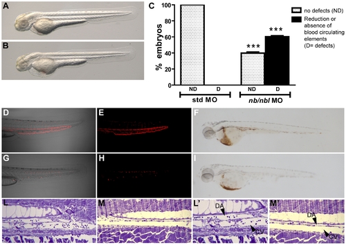
The nb/nbl knockdown affects erythrocytes development. A–B. The general morphology of 48 hpf nb/nbl morphants is substantially unaffected (B) when compared to control embryos (A). C. Hematopoietic defects in nb/nbl morphants at 48 hpf: ∼60% of the nb/nbl morphants displayed a severe hematopoietic defect (n = 191). 100% of control embryos was unaffected (n = 93). *** p<0.001 vs std MO. D–I. Detailed images of the blood flow in the trunk-tail region of 48 hpf Tg(gata1:dsRed) embryos injected with std MO (0.8 pmol/embryo; D, E) and nb/nbl MO (0.8 pmol/embryo; G, H). In nb/nbl morphants no circulating red cells are detectable within the trunk vasculature. Whole embryo o-dianisidine staining on 48 hpf std MO embryos (F) and nb/nbl morphants (I). L–M′. Longitudinal semithin plastic sections of std MO (L, L′) and nb/nbl MO-injected embryos (M, M′) at 2 dpf. Fewer elements are detectable within the Dorsal Aorta (DA) and the Caudal Vein (CV) in nb/nbl morphants when compared to control embryos. L′, M′: higher magnification of L, M. |

Expression of hematopoietic genes in nb/nbl morphants. A–D. Flat mounted 10-ss embryos. Triple WISH were performed on std MO (A, C) and nb/nbl MO-injected embryos (B, D). The expression of myoD in somitic and pre-somitic mesoderm appeared unaltered in nb/nbl MO embryos (B, D). The expression of krox20 in the rombomeres 3 and 5 is unaffected in nb/nbl morphants (B, D). A–B. The expression of scl appears reduced in the posterior PLM (black arrowheads). C–D. gata1 expression is reduced in nb/nbl morphants, particularly in the posterior PLM (black arrowheads). E–N. Lateral view of 22–24 hpf std MO embryos (E, G, I, M) and nb/nbl morphants (F, H, L, N). In nb/nbl morphants at 22 hpf gata1 expression is reduced in the ICM (F); ikaros and βe1 globin are also downregulated at 24 hpf (H, L). The myeloid marker pu.1 is expressed at normal levels in nb/nbl MO-injected embryos (N). EXPRESSION / LABELING:
|

dsRed+ erythroid cells are dramatically reduced at 28–30 hpf. A–H. Tg(gata1:dsRed) embryos injected with std MO (A, B, E, F) and nb/nbl MO (C, D, G, H) were examined by confocal microscopy between 24–30 hpf. Fluorescent images (B, D, F, H) were merged with bright field images (A, C, E, G). In nb/nbl morphants at 24–26 hpf red fluorescent erythroid cells are present within the ICM (C, D) but at 28–30 hpf the overall fluorescence of nb/nbl morphants appears strongly reduced (G, H). I–N. Whole-mount double immunofluorescence on Tg(gata1:dsRed) nb/nbl morphants at 28–30 hpf to detect caspase-3 activation (green) and DsRed (Red). Single optical section of nb/nbl morphants obtained by confocal microscopy (20x magnification, I). I. White spots, indicating double positive cells, have been pseudocoloured according to the region of interest (ROI1 in O). The sub-image area, shown in detail in panels L–N, is highlighted by the white box. L–N. Insets of single channel fluorescent images (L, M) and merge (N) are shown. Activated caspase-3 overlaps with some dsRed+ cells (white arrowheads, L). O. Fluorogram shows the degree of colocalization between red signals (dsRed) and green signals (activated caspase-3); colocalization is indicated by the region of interest (ROI1). |

Erythroblasts fail to differentiate into erythrocytes in apparently unaffected nb/nbl morphants. A–B. Bright-field microscopy of blood cells in the caudal arteries of living 48 hpf std MO embryos (A) and nb/nbl morphants (B). nb/nbl MO-injected embryos with no apparent hematopoietic defects show the presence of abnormal-shaped blood cells (white arrowheads) in the blood flow (B). C–D, G–H. Wright-Giemsa staining of circulating embryonic red blood cells from controls and nb/nbl morphants at 52 hpf (C, D) and 3 dpf (G, H). Erythroid cells of apparently unaffected nb/nbl MO injected embryos were larger, showed a large nucleus and had more basophilic cytoplasm indicating the presence of maturation defects (D, H). Asterisks indicate representative erythroid cells with maturation defects. E–F. WISH using gata1 on 48 hpf std MO (E) and nb/nbl MO-injected embryos (F). gata1 expression persists in red blood cells of nb/nbl morphants revealing the presence of immature red cells (black arrowhead). |

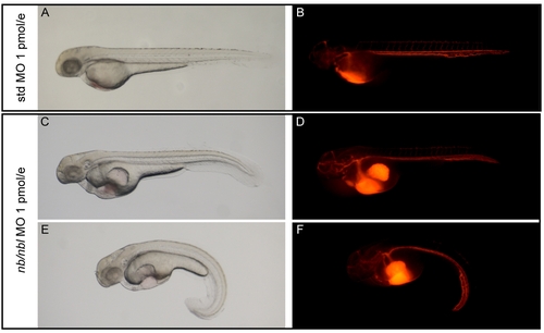
Dose dependent hematopoietic phenotype induced by nb/nbl MO. A–D. Tg(gata1:dsRed) std MO and nb/nbl MO (0.8pmol/embryo) at 48 hpf. Images were taken in bright field (A, B) and using a rhodamine emission filter (C, D). E–G. Analysis of the hemoglobin content by whole embryo o-dianisidine staining. 48 hpf std MO embryos (E) and nb/nbl morphants (1 pmol/embryo; F, G). At this dose the 62% of nb/nbl morphants shows a drastic reduction of the hemoglobin content (F), an additional 30% shows complete loss of hemoglobin staining (G). H. Injection of different doses of nb/nbl MO (0.8–1 pmol/embryo) produces a dose-dependent hematopoietic phenotype. The data are referred to a single typical experiment. |
The heart functionality and the axial vasculature are not drastically compromised in nb/nbl morphants. Microangiography experiments, were performed on 2 dpf controls (A, B) and embryos injected with nb/nbl MO at the high dose of 1 pmol/embryo (C–F). In nb/nbl morphants (D, F) the injected dye flows into the main axial vessels as in control embryos (B). |
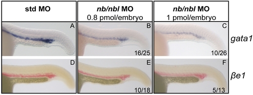
Dose dependent reduction of gata1 and βe1 globin expression in nb/nbl morphants. WISH were performed on controls (std MO; A, D) and embryos injected with different doses of nb/nbl MO (0.8 pmol/embryo, B, E; 1 pmol/embryos, C, F). In the ICM of 22–24 hpf nb/nbl MO-injected embryos the downregulation of gata1 and βe1 appears dose dependent. |
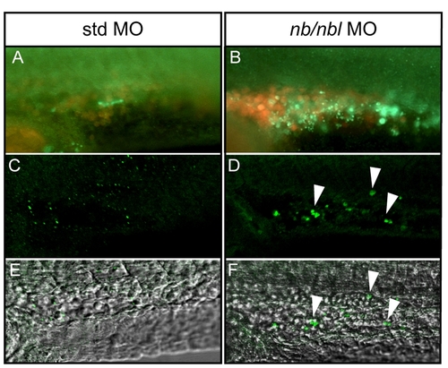
Caspase-3 activation in nb/nbl morphants at 28–30 hpf. A–B. Whole-mount immunofluorescence to detect caspase-3 activation (green signal), detailed view of the ICM region of 26–28 hpf Tg(gata1:dsRed) embryos injected with std MO (A) and nb/nbl MO (B). C–F. Single optical sections of 26–28 hpf control and nb/nbl MO-injected embryos in which whole-mount immunofluorescence for caspase-3 activation (green signal) was performed. Fluorescent images (C, D) were merged with bright field images (E, F). Detailed view of the ICM region. Caspase-3 activation can be detected in erythroid cells of nb/nbl morphants (white arrowheads; D, F). |

Single numb and numblike knockdown reproduce the nb/nbl morphants hematopoietic defects with low penetrance. A–B. Injection of nb MO1 and nbl MO1 specifically blocks splicing of the targeted pre-mRNAs. PCR reactions were performed on cDNAs retrotranscribed from total RNA extracted from 29 hpf nb MO1 injected embryos (1.4 pmol/embryo; A), nbl MO1 injected embryos (0.3 pmol/embryo; B), std MO injected embryos (0.3 pmol/embryo or 1.4 pmol/embryo; A, B). β-actin has been tested as an internal control (data not shown). A control PCR reaction performed without cDNA is shown in lane 3 of both the boxes (A, B). Primers: nb MO1-5′: CACCAGTGGCAGACCGATGAA nb MO1-3′: ACCGCTCGCACAGCCTTCTTA nbl MO1-5′: TCGGGCTGGTGGAGGTGGAT nbl MO1-3′: CCGTCACGGCAGATGTAAGAG. C. Single injection of nb MO1 (1.4 pmol/embryo) or nbl MO1 (0.3 pmol/embryo) in Tg(gata1:dsRed) produces the hematopoietic phenotype respectively in ∼19% (n = 99) and ∼25% (n = 125) of the MO injected embryos (*p<0.05 vs std MO no defects, **p<0.01 vs std MO no defects, #p<0.05 vs std MO defects, ##p<0.01 vs std MO defects). 100% of control embryos was unaffected (n = 65). PHENOTYPE:
|

her6 is ectopically expressed in nb/nbl morphants. A–B. Posterior view of 8–10-ss embryos. The nb/nbl morphants display an enlarged expression domain of the Notch target gene her6 within the PLM region (B; black arrowheads), when compared to controls (A). C. Percentages of rescue of the hematopoietic phenotype in nb/nbl morphants treated with different concentrations of DAPT. |

Unillustrated author statements |