
- Title
-
Funduscopy in adult zebrafish and its application to isolate mutant strains with ocular defects
- Authors
- Tschopp, M., Takamiya, M., Cerveny, K.L., Gestri, G., Biehlmaier, O., Wilson, S.W., Strähle, U., and Neuhauss, S.C.
- Source
- Full text @ PLoS One

Images of zebrafish eyes. (A) Overview of anaesthetized fish with cover glass, (B) shows a normal, transparent lens, (C–F) examples of opaque lenses (cataracts): (C) small inclusion are present in an otherwise normal lens, (D) massive cataract, (E) including cracks in the lens, and (F) membranous cataract. Scale bar: 500 μm. PHENOTYPE:
|
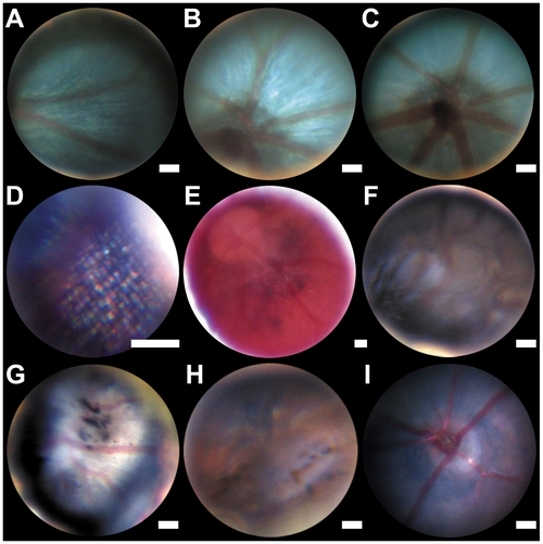
Zebrafish and mouse fundus images. (A–D) wild type zebrafish fundus at the level of the mid-peripheral retina (A), optic disc (B and C) and photoreceptor level, showing the presumed photoreceptor mosaic (D). Hypopigmented fundus of the zebrafish albino strain (E). Tortuous arteries and a darkened retina (F), black spots are visible in the retina (G and H). Fundus Image of a wild type mouse (I). Estimated scale bar: 50 μm. PHENOTYPE:
|
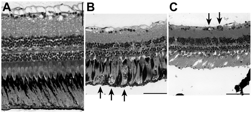
Adult retina sections. (A) Wild type zebrafish. (B) Right eye and (C) left eye of a fish with black spots in the retina; the funduscopy of this fish is shown in Figure 3 (H). Arrows indicate cells with black granules: likely macrophages filled with RPE cells or detached RPE cells. The detached RPE in (C) is a histological artefact. Scale bar: 50 μm. PHENOTYPE:
|