
- Title
-
Carboxypeptidase A6 in zebrafish development and implications for VIth cranial nerve pathfinding
- Authors
- Lyons, P.J., Ma, L.H., Baker, R., and Fricker, L.D.
- Source
- Full text @ PLoS One
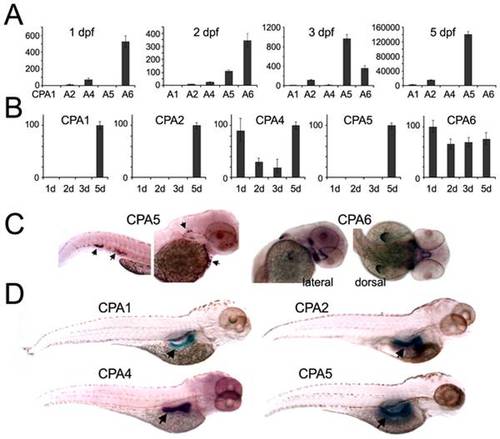
Temporal and spatial expression of zebrafish CPA genes. (A, B) qPCR was performed using cDNA prepared from zebrafish at the indicated developmental time points. All values were normalized to β-actin, and are shown in (A) as expression relative to CPA1 mRNA at 2 dpf, or in (B) as expression of each gene relative to its highest level (100%). (C, D) In situ hybridization was used to determine the spatial distribution of CPA mRNA expression. (C) At 2 dpf CPA5 mRNA (arrows) was detected in a mast cell population and CPA6 mRNA was seen in precursor tissues found in the stomodeum, posterior to the eyes, and in the pectoral fin buds. (D) At 4 dpf, mRNA for CPA1, 2, 4, and 5 were expressed in the pancreas. |

Detailed analysis of CPA6 mRNA expression throughout zebrafish development. In situ hybridization indicated CPA6 mRNA (purple) is found in newly formed somites (A–D), ectodermal cells of the tail (C,D), a tissue posterior to the eye (E–H, arrows), the stomodeum (G, asterisk), and the pectoral fins (H, arrowhead). CPA6 expression posterior to both left and right eyes can be seen in F and H. A summary of the spatial (I) and temporal (J) expression of CPA6 throughout zebrafish development is shown. EXPRESSION / LABELING:
|

Distribution of CPA6 mRNA compared with tissue-specific markers at 2 dpf. (A) A general marker of muscle precursors, myogenin [16], labels most extraocular muscles (orange) by in situ hybridization, but does not co-localize with CPA6 mRNA (purple). (B) Myogenin mRNA, as well as MyoD mRNA, is also found in the pectoral musculature, unlike CPA6 mRNA which is ectodermal. CPA6 mRNA does not co-localize with Meox1 (C), or Lbx1 (D), both putative markers of the lateral rectus muscle in the chick, but likely not in the zebrafish. (E) A schematic of a 2 dpf zebrafish indicates the relative spatial expression of CPA6, myogenin (in lateral rectus), Lbx1 and Meox1. lr, lateral rectus; mr, medial rectus; sr, superior rectus; ah, adductor hyoideus; cd, constrictor dorsalis; MD, myodome precursors; O.V., otic vesicle. EXPRESSION / LABELING:
|
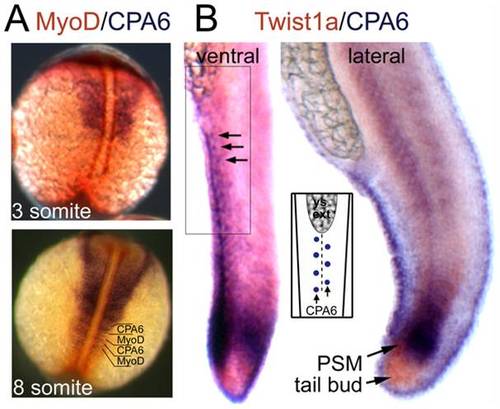
Distribution of CPA6 mRNA compared with somitogenesis markers. In situ hybridization was performed with RNA probes specific for (A) CPA6 (purple) and MyoD (orange) at 3 and 8 somite stages (11 and 14 hpf), and (B) CPA6 (purple) and Twist1b (orange) at 24 hpf. Arrows indicate ectodermal cells arranged along the ventral ridge of the tail and expressing CPA6. The regular arrangement of these cells, also found along the dorsal ridge, is illustrated in the inset. PSM, presomitic mesoderm; ys ext, yolk-sac extension. EXPRESSION / LABELING:
|

Knockdown of CPA6 gene function. (A) Morpholinos were designed to interfere with normal CPA6 mRNA splicing (Ctr-MO: control morpholino; MO-2 and MO-3: CPA6 specific morpholinos; lines: MO binding sites; arrows: primer binding sites). (B) RT-PCR using RNA extracted from either uninjected or morpholino-injected embryos (3 ng) showed CPA6-specific knockdown. (C) 48 hpf embryos injected with control morpholino and CPA6 MO-2. CPA6 morphants exhibited yolk-sac edema. (D) DiI injected into the lateral rectus muscle (LR) of 8 dpf Ctr-MO and MO-2 CPA6-morphant zebrafish larvae retrogradely labeled the VIth cranial nerve (nVI). Scale bar = 40 µm. PHENOTYPE:
|

Optokinetic reflex measurement of CPA6 morphants at 5 dpf. (A) Morphology of CPA6 morphants. (B) Schematic illustration of central neural pathways for either lateral or medial rectus mediated optokinetic tracking and saccades. (C) Representative example of eye position (blue trace; top panel), fast phase saccades (green hatching; top panel) and slow phase tracking velocity (cyan trace; bottom panel) in response to ±20°/s velocity steps optokinetic stimuli (grey trace; bottom panel) at 0.0625 Hz. (D) Quantification of saccadic velocity produced by either the lateral (orange bars) or medial rectus muscle (grey bars) in CPA6 morphants and controls. Data are presented as mean ± S.D. (E) Representative eye tracking, saccades and gain in a MO-2 (3 ng) morphant and corresponding control. (F) Optokinetic reflex in MO-3 (6 ng) morphants and corresponding control. PHENOTYPE:
|