
- Title
-
A novel transgenic zebrafish model for blood-brain and blood-retinal barrier development
- Authors
- Xie, J., Farage, E., Sugimoto, M., and Anand-Apte, B.
- Source
- Full text @ BMC Dev. Biol.

Development of the BRB and BBB in zebrafish. FD4 (4,000 Da, green) was injected to the sinus venosus of Tg(flk1:mCherry) embryos at 2 dpf (A, B, G, H), 2.5 dpf (C, D, I, J) and 3 dpf (E, F, K. L). Live confocal images of the hyaloid vessels (A-F, side view), brain vessels (G-J, dorsal view) and trunk vessels (K-L, side view) were obtained from 10 to 150 minutes after injection. Insets and panel L are images of established vasculature of the injected embryos (red). In the brain, boundaries of the middle mesencephalic central artery (MMCtA) are indicated by arrows in G-J, cerebellar central artery (CCtA), by blue arrows in G-J and posterior mesencephalic central artery (PMCtA) by asterisks in I&J. Arrows in K&L indicate intersegmental vessels and arrowhead in K shows the myotomal boundaries. A small molecule, fluorescein (376 Da, green), was also used as a tracer in the injection assay (M-R). In the 2.5 dpf embryos (M&N), the PMCtA (asterisk) could not be differentiated, and boundaries of MMCtA (arrows) and CCtA (blue arrows) were enlarged and became blurred 30 minutes after injection. However in the 3 dpf embryos injected with fluorescein (O-R), boundaries of the MMCtA (arrows), PMCtA (asterisk), CCtA (blue arrows) and the hyaloid vessels (Q&R) remained sharp and clear after 50 minutes of injection. The leaked FD4 accumulated mostly in brain ventricles (arrowheads in G-J). In contrast, most of the leaked fluorescein did not accumulated in the brain ventricle (arrowheads in M&N), but evenly diffused throughout the brain (M-P). Scale bars: 50 μm. |

Spatial and temporal expression of claudin-5 in the developing BBB. Tg(flk1:EGFP) embryos at 2 dpf, 2.5 dpf, and 3 dpf were stained with a monoclonal claudin-5 antibody. Confocal images of whole mount embryos were analyzed for claudin-5 expression (red) (A-E and H-M; Alexa Fluor 568 labeled secondary antibody) in developing blood vessels (green) (A′-E′ and H′-M′; EGFP labeled vascular endothelial cells; F&G, merged pictures). All the samples are oriented with anterior to the left. A-E and A′-E′, lateral views; other panels, dorsal views. Scale bars: 50 μm. EXPRESSION / LABELING:
|

Spatial and temporal expression of claudin-5 in the developing BRB. Tg(flk1:EGFP) embryos and larvae from 2 dpf to 4 dpf were stained with mouse anti-claudin-5. Confocal images of whole mount were analyzed for claudin-5 (red) (A-I; Alexa Fluor 568) expression in hyaloid blood vessels (green) (A′-D′ and G′-I′; EGFP). All the panels are dorsal views except G&G′. The claudin-5 signal in the hyaloid vessels at 2 dpf (A&A′) is minimal. At 2.5 dpf (B&B′), and 3 dpf (C&C′) the staining in the hyaloid vasculature is increased. Claudin-5 is also expressed in the hyaloid artery (asterisks) and outer limiting membrane of the retina (shaded arrow) and inner plexiform layer (shaded arrowhead). (D&D′) At 3.5 dpf, the wider cone-shape staining is lost and the claudin-5 signal overlaps completely with the HA (asterisks). The vessel connecting the HV and the inner optic circle (IOC) also has a strong claudin-5 signal (blue arrows in D&D′, G&G′). (E&F) In the retina, the claudin-5 signal does not clearly outline the polygonal RPE (arrow) until 3 dpf. The insert is an enlarged view of the dashed square. At 4 dpf (G-I), the HV (arrows in G&G′, H&H′) and the HA, but not the choroidal vasculature (such as the IOC, indicated by arrowhead in G′), express claudin-5. The claudin-5 is also expressed around the foramen (opening) through which the HA penetrates the retina (arrow in I). The panel J is a schematic illustrating expression of claudin-5 in optic vasculature of zebrafish. Scale bars: 50 μm. |

Claudin-5a and 5b expression in hyaloid vasculature. (A) The 3′ ends of three zebrafish claudin genes have significant homology with the C-terminal of mouse claudin-5. Claudin-5a (zgc 85723; GenBank: NM_213274) and claudin-5b (zgc 103419; GenBank: NM_001006044) are mostly homologous to claudin-5a of Fugu rubripes, with 82% identical (plus 8% similar) and 75% identical (plus 14% similar) amino acid sequences, respectively. The zebrafish claudin-h (GenBank: NM_131767) is mostly homologous to claudin-3a of Fugu. (B-E′) Whole mount in situ hybridization of claudin-5a expression. (B) Claudin-5a is expressed in the CNS (midbrain, hindbrain, ventricular zone, and epiphysis) at 1 dpf. (C-E & C′-E′) From 1.5 to 2.5 dpf, claudin-5mRNA is detected in the hyaloid vasculature (black arrows), hyaloid artery (white arrows) and the cornea (arrow heads). (F-H) Claudin-5b is expressed in the entire vascular system at 1 dpf (arrow and arrowheads in F), and is confined to the blood vessels of the brain (arrows in G) and cardiovascular system (arrowhead in G) at 2 dpf. Similar to claudin-5a, expression of claudin-5b in the hyaloid vasculature (black arrow in H) and hyaloid artery (white arrow in H) is seen at 1.5 dpf and lasts till 3 dpf. B-E, dorsal view; C′-E′, side view; F-H, side view. |

Expression pattern of DBP-EGFP in the blood plasma of l-fabp:DBP-EGFP transgenic zebrafish. Images were obtained from Tg(l-fabp:DBP-EGFP;flk1:mCherry) fish to visualize DBP-EGFP (green) and endothelial cells lining blood vessels (red)(mCherry). The fish were oriented with anterior to the left. All panels are side views except E. (A) green fluorescence in eggs laid by female Tg(l-fabp:DBP-EGFP) fish. (B) DBP-EGFP expression at 1.5 dpf. (C, C′, D & E) DBP-EGFP expression at 2 dpf. The central arteries (arrows) are the first distinguishable blood vessels. (F, F′, G & H) DBP-EGFP expression at 60 hpf. The dashed rectangles in H and K are merged pictures of red and green channels. The branchial arches (blue arrow) can be differentiated at the time, but boundaries of HV are still not clear (arrowhead). At 3 dpf (I, I′, J & K), the boundaries of the HV become sharp (arrows in I&I′). More vessels appear in the liver (dashed rectangle) and more brain vessels can be seen (arrows in G and J). At 4 dpf (L, L′, M & N), the EGFP-infused hyaloid and brain vessels can be easily differentiated from the fluorescent background (arrows in L, L′ and M), as can the intersegmental vessels (arrow in N) and the dorsal aorta (blue arrow). Accumulation of DBP-EGFP in the myotomal boundaries (arrowhead) (N) is observed from leakage out of the vasculature. At 5 dpf (O), the HV (blue arrow) and the trunk vessels (arrow) are distinguishable, but the brain vessels are not (arrowhead), due to the increased fluorescent background in the brain. (P) At 60 dpf, the HV is still distinguishable. EXPRESSION / LABELING:
|
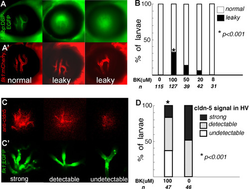
Bradykinin mediated disruption of zebrafish BRB (A, A′ & B). The l-fabp:DBP-EGFP;flk1:mCherry double transgenic larvae were treated with 8 to 100 μM BK from 5 to 9 dpf. (A) DBP-EGFP (green) in hyaloid vessels of control (left panel), leaky hyaloid vessels (middle and right panel); (A′) endothelial lining of blood vessels, mCherry (red). (B) At 9 dpf, the larvae were scored for the presence of leaky hyaloid vessels. Both Fisher′s test and chi-square test indicate that the BK treatment results in significantly increased numbers of larvae showing GFP leakage compared with those treated with control buffer (P < 0.001). (C) Claudin-5 expression (red) was evaluated by whole mount immunohistochemistry in Tg(flk1:EGFP) larvae exposed to 100 μM BK and scored as strong expression (left panel), detectable (middle panel) and undetectable (right panel); (C′) endothelial lining of blood vessels, flk1:EGFP (green). (D) Approximately 47 BK treated and 46 untreated flk1:EGFP larvae were evaluated for claudin-5 expression in the hyaloid vasculature. EXPRESSION / LABELING:
|

Expression of ZO-1 in the developing BRB and BBB of zebrafish. Tg(flk1:EGFP) embryos at 2 dpf to 3 dpf were stained with rabbit anti-ZO-1; confocal images were analyzed for ZO-1 expression (red) (A-G; Alexa Fluor 568) and blood vessels (green) (A′-E′; EGFP). A&A′, lateral views; the other panels, dorsal views. (A&A′) The ZO-1 signal is high in the gut (shaded arrow) and low in the intersegmental vessels (arrows). (B&B′) At 2 dpf, ZO-1 is localized to the HV (arrows) and the HA (arrowheads). (C&C′) At 3 dpf, the HV (arrows), inner plexiform layer (shaded arrowhead) as well as the outer limiting membrane (shaded arrow) show a strong signal of ZO-1. (D&D′, E&E′) At 2 dpf, most brain vessels express ZO-1, including the BCA (arrowheads). Similar to the claudin-5 antibody, the ZO-1 antibody binds to many non-endothelial structures in the brain (shaded arrows and shaded arrowhead) besides the brain vasculature (arrows). (F&G) At 2 dpf, the ZO-1 antibody can stain the polygonal RPE cells (arrows) clearly. The insert in G is an enlarged view of the dashed square. Scale bars: 50 μm. EXPRESSION / LABELING:
|

In situ hybridization of cldn-5a, cldn-5b and cldn-h. Wild type embryos at 2.5 dpf were probed with anti-sense probes of cldn5a, cldn5b and cldn5h. The cldn-5a and cldn-5b had very strong signal in the hyaloid vasculature around the lens, while the cldn-h did not. Two sense controls did not show any signal. EXPRESSION / LABELING:
|

Claudin 5b is expressed in hyaloid vessels. A 5.6 kb upstream sequence of claudin-5b can drive expression of EGFP in the hyaloid vessels. The inset is an enlarged view of the green hyaloid vessels. |

Bradykinin induces BRB breakdown. At 5 dpf, 20 double transgenic (l-fabp:DBP-EGFP;flk1:mCherry) larvae were injected with 2 μL of 20 μM bradykinin in the right eye (B, B′, C and C′). 3 hours after injection, the BRB was found to be disrupted in 18 injected eyes, as indicated by the leakage of DBP-EGFP (green) (B, C) from the hyaloid vessels (red) (B′C′) which showed normal morphology. No leakage was observed in any of the BSA injected contra-lateral negative control eyes (A, A′). |

DBP-EFGP is a 78 kDa protein in l-fabp:DBP-EGFP zebrafish. The expression level and molecular weight of DBP-EGFP was analyzed by western blot analysis of lysates from l-fabp:DBP-EGFP (A) and wild type (B) embryos using anti-GFP antibodies (Abcam, ab290). A 78 kDa protein was detected in lysates from transgenic embryos. |
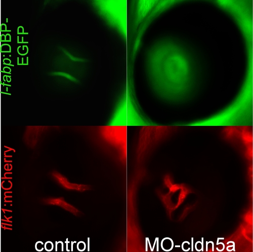
Morpholino knockdown of claudin 5a in zebrafish embryos results in BRB breakdown. Cldn-5a was knocked down by injecting morpholino-cldn5a into double transgenic l-fabp:DBP-EGFP;flk1:mCherry embryos at the 2-cell stage. At 4 dpf, leakage of DBP-EGFP(green) from the hyaloid vasculature (red) was observed. A 5 bp-mismatched Morpholino was injected as control and showed an intact BRB. PHENOTYPE:
|