
- Title
-
SCORE imaging: specimen in a corrected optical rotational enclosure
- Authors
- Petzold, A.M., Bedell, V.M., Boczek, N.J., Essner, J.J., Balciunas, D., Clark, K.J., and Ekker, S.C.
- Source
- Full text @ Zebrafish
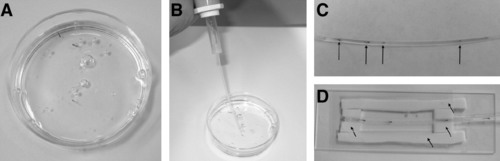
SCORE tools. (A) Anesthetized larval zebrafish are placed into a methylcellulose (pictured) or low-melting-point agarose solution before being loaded into the capillary housing. (B) A pipette pump with capillary adaptor is used to facilitate loading of zebrafish into the capillary housing. (C) Multiple larvae (arrows) can be imaged in a single capillary, allowing for rapid screening of mutants. (D) A corrective solution (water pictured) is placed upon the capillary to reduce distortion. Foam barriers, marked by arrows, are placed upon the glass slide to help contain corrective solution and to provide a friction surface for ease of rotation. |

Specimen in a Corrected Optical Rotational Enclosure (SCORE) imaging of living animals. Zebrafish imaging is facilitated by use of a fluoro-carbon tube for housing. (A) Zebrafish images taken in polymer tubing produce a distorted image (arrows) due to the change in refractive index between the tubing and the air. Cartoon images representing a fish immobilized using traditional techniques on a skewed plane (B), a larval fish within a polymer tube without a corrective solution producing distortion (C), and within a corrective solution to produce a clear image (D). Fluorinated ethylene propylene (FEP) tubing produces a clear image at 50x (E ) (also see Supplemental Movie S1, available online at www.liebertonline.com), 100x (F), and 200x (G) of a 5 day postfertilization (dpf) larvae. A 5 dpf Tg(gata-1:DsRed)sd2 larvae in FEP tubing produces a clear background-free image at 50x (H) (also see Supplemental Movie S2, available online at www.liebertonline.com) and 100x (I). A 5 dpf Tg(fli:eGFP)y1 in FEP tubing produces a clear background-free image at 50x (J) (also see Supplemental Movie S3, available online at www.liebertonline.com) and 100x (K). |
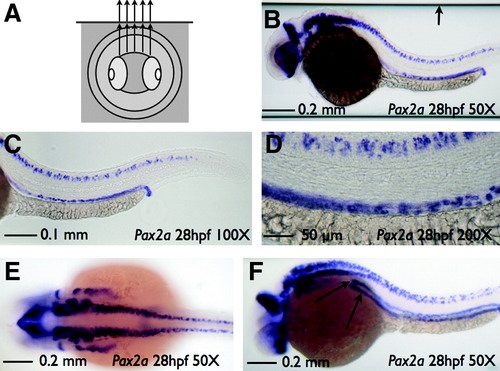
SCORE imaging of a fixed specimen. Imaging of a pax2a whole-mount in situ hybridization embryo and capillary housing in 75% glycerol. (A) Cartoon depicting the use of glycerol both inside and outside of the glass tube producing an undistorted image of a fixed embryo. (B) Sagittal image of embryo with pax2 staining at 50x magnification shows no distortion (also see Supplemental Movie S4, available online at www.liebertonline.com). Note the edges of the capillary (arrow) that can be readily cropped for publication presentation. Sagittal embryo image of pax2 staining at 100x (C) or 200x (D) magnification shows no distortion. (E) Coronal image of embryo at 50x magnification. (F) Angled image of pax2 staining at 50x magnification. Rotation is angled slightly (∼30°) to show a more detailed view of kidney tubule staining (arrows). |
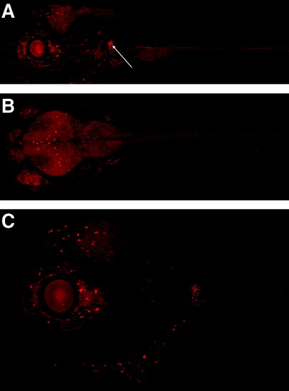
SCORE imaging of fluorescent, living zebrafish. Imaging SEC0124 of a 5 dpf mRFP-labeled protein trap zebrafish housed in FEP tubing. (A) Sagittal image of an mRFP-labeled protein trap zebrafish shows enhancement in specific locations in the neural region as well as a bright kidney (Arrow). (B) Coronal image of an mRFP-labeled protein trap zebrafish includes ubiquitous neural expression with enhancement in specific locations. (C) Sagittal image of an mRFP-labeled protein trap zebrafish shows a general neural haze with enhancement in specific locations at 100x magnification (also see Supplemental Movie S5 [showing the spatial location of RFP patterning], available online at www.liebertonline.com). EXPRESSION / LABELING:
|