
- Title
-
Role of G protein-coupled estrogen receptor 1, GPER, in inhibition of oocyte maturation by endogenous estrogens in zebrafish
- Authors
- Pang, Y., and Thomas, P.
- Source
- Full text @ Dev. Biol.
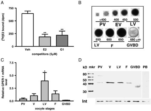
Estrogen binding to ovarian membranes and pattern of GPER mRNA and protein expression in zebrafish oocytes at different developmental stages. (A) Single-point competitive binding of [3H]-E2 to ovarian plasma membranes in the presence of 500-fold excess E2 and G-1 competitors. ∗∗, P < 0.01 vs. vehicle (Veh). (B) Appearance and diameters of zebrafish oocytes at different developmental stages. PV, pre-vitellogenic oocytes. EV, early-vitellogenic oocytes. LV, late-vitellogenic oocytes. F, full-grown oocytes. GVBD, oocytes undergoing germinal vesicle breakdown. (C) Quantitative RT-PCR measurement of GPER mRNA levels in the oocytes at different developmental stages. (D) Western blot analysis of GPER protein expression on the plasma membranes of oocytes at different development stages using a specific GPER antibody. mkr, protein size marker. Int, integrin loading control. PB, peptide block. |

Localization of GPER in zebrafish ovarian tissue cryosection samples by immunohistochemistry using a specific GPER antibody. (A–D) Early-vitellogenic stage follicles. (E–H) Late-vitellogenic stage follicles. (A, E) Green fluorescent staining of zebrafish GPER on oocyte plasma membranes (om). (B, F) DAPI staining of nuclear DNA of follicular cells (f). (C, G) Merge. (D, H) Detection of GPER with the GPER antibody that had been neutralized with the antigen peptide and merged with DAPI stained images. Scale bar = 100 μm. EXPRESSION / LABELING:
|

Effects of enzymatic and manual removal of ovarian follicle cells (defolliculation) on oocyte maturation. (A) Intact ovarian follicles stained with DAPI, before and after collagenase treatment. int, intact follicle; df, defolliculated follicles. (B) Effects of E2 (100 nM) on the GVBD of intact (int) and enzymatically defolliculated (df) oocytes after 6 h of incubation. (C) Effects of 100 nM E2 and G-1 on GVBD of manually defolliculated oocytes. ab, cd, ef: P < 0.05. df, manually defolliculated oocytes. (N = 3). |
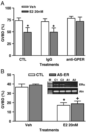
Involvement of GPER in zebrafish oocyte maturation. (A) Effect of E2 on GVBD of defolliculated full-grown oocytes treated with IgG and fish GPER antibody (1:300). ∗, P < 0.05 compared to Veh. (B) Effect of E2 (20 nM) on GVBD of LV oocytes (∼ 550 μm) that had been micro-injected with control (CTL) and ERα antisense morpholino oligos. AS-ER, antisense ERα oligos. ∗, +, P < 0.05 compared to vehicle (Veh). Gel image, RT-PCR detection of ERα mRNA levels in the micro-injected oocytes. M, DNA size marker. C1 and C2, control 1 and 2. A1 and A2, antisense oligos. Atn, actin control. (N = 3). |

RT-PCR detection of ER expression in zebrafish oocytes. |
Reprinted from Developmental Biology, 342(2), Pang, Y., and Thomas, P., Role of G protein-coupled estrogen receptor 1, GPER, in inhibition of oocyte maturation by endogenous estrogens in zebrafish, 194-206, Copyright (2010) with permission from Elsevier. Full text @ Dev. Biol.