FIGURE SUMMARY
Title

Broad-Minded Links Cell Cycle-Related Kinase to Cilia Assembly and Hedgehog Signal Transduction

Authors
Ko, H.W., Norman, R.X., Tran, J., Fuller, K.P., Fukuda, M., and Eggenschwiler, J.T.
Source
Full text @ Dev. Cell

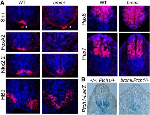
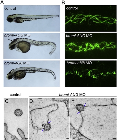
Neural Patterning in bromi Mutants
(A) Spinal neural tubes of E10.5 wild-type (WT) and bromi mutants immunostained for markers of neural identity (in red, DAPI in blue). bromi mutant neural tubes lacked the floor plate, marked by Shh and FoxA2, although the notochord (arrowheads) expressed Shh normally. Nkx2.2+ p3 cells were inappropriately positioned in the mutant ventral midline and reduced in number. The Pax6+ and HB9+ (motor neuron) domains, which are supported by intermediate, but not high, levels of Shh, ectopically expanded into the most ventral regions in the mutants. The ventral limit of the Pax7+ domain, set by low levels of Shh signaling, was not affected in bromi mutants.
(B) X-gal staining of E10.5 neural tubes from WT and bromi embryos heterozygous for the Ptch1-LacZ insertion. Ptch1-LacZ is expressed in a ventral-to-dorsal gradient in response to Shh. Expression in the ventral one-third of the bromi mutant neural tubes (n = 3) was consistently weaker compared with that of WT.

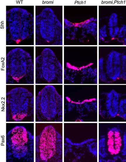
bromi Is Epistatic to Ptch1
Spinal neural tubes from E9.5 WT, bromi, Ptch1, and bromi,Ptch1 mutant embryos immunostained for markers of neural fate. In Ptch1 mutants, Shh, FoxA2, and Nkx2.2 were broadly expressed throughout the open neural tubes, and Pax6 expression was absent. The Ptch1 mutant phenotype was strongly suppressed in bromi,Ptch1 double mutants, whose the closed neural tubes were small, yet showed expression patterns similar to bromi single mutants.

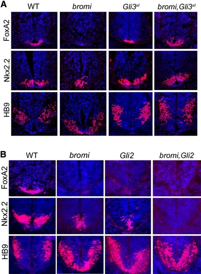
Patterning in bromi,Gli3xt and bromi,Gli2 Double Mutants
(A) FoxA2, Nkx2.2, and HB9 immunostaining of E10.5 WT, bromi, Gli3xt, and bromi,Gli3xt double-mutant spinal neural tubes. Expression of these markers was normal in Gli3xt mutants, but disruption of Gli3 restored floor plate specification in bromi,Gli3xt double mutants.
(B) FoxA2, Nkx2.2, and HB9 immunostaining of E10.5 WT, bromi, Gli2, and bromi,Gli2 double-mutant spinal neural tubes. Ventral neural patterning is similar in bromi and Gli2 single mutants. bromi,Gli2 double-mutant neural tubes lacked Nkx2.2+ cells, whereas they were always detected, albeit in reduced numbers, in either single mutant.

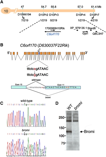
Positional Cloning of bromi
(A) bromi was mapped to a 2.3 Mb region on mouse chromosome 10 by analysis of recombinants (recombination events, out of 219 opportunities scored for each marker, are listed). This interval contained six known and predicted genes, which were fully sequenced in bromi and parental (C57BL/6J) strains. (B and C) An A-to-G splice acceptor mutation at the intron 10/exon 11 junction of C6orf170 was identified in bromi samples (trace shown in [C]), and sequencing of RT-PCR products showed that bromi mutants use a cryptic splice acceptor site 13 base pairs downstream.
(D) Western blotting of WT and bromi mutant embryonic extracts with polyclonal antisera against the Bromi protein.

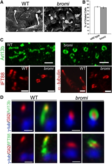
Cilia Defects in bromi Mutants
(A) Neuroepithelial cilia from E10.5 WT and bromi mutant neural tubes analyzed by scanning electron microscopy. bromi mutant cilia showed a swollen or bulbous morphology (arrowheads). Scale bars, 1 μm.
(B) Cilia frequency in the WT and bromi mutant neuroepithelium. Error bars represent ± 1 SEM.
(C) Confocal images of neural tube cilia stained with Arl13b (upper panels), IFT88 and acetylated α-tubulin (lower panels). Scale bars, 1 μm.
(D) Neural tube cilia from E10.5 WT and bromi mutant embryos immunostained for γ-tubulin/Arl13b/ Gli2 or γ-tubulin/IFT88/Gli2. Scale bars, 0.5 μm.

bromi Regulates the Association Between Ciliary Membranes and Axonemes
(A) Zebrafish embryos (72 hpf) injected with MOs against the start site (bromi- AUG) and exon 8/intron 8 splice site (bromi-e8i8) of the zebrafish bromi homolog resulted in abnormal tail curvature. bromi-AUG MO fish exhibited hydrocephalus (arrow), while this was less frequent in bromi-e8i8 MO fish.
(B) Confocal microscopy of acetylated α-tubulin-stained cilia in the posterior (distal early/late) regions of the zebrafish kidney tubules. Ciliary morphology in the anterior (proximal convoluted/straight) regions of the tubules was not appreciably affected in the morphants.
(C–E) Posterior pronephric cilia visualized by TEM showing 9 + 2 axonemal structure in 48 hpf control and bromi-AUG MO fish. The membranes of control cilia were tightly associated with axonemes (C), whereas they detached along one side of the axonemes (blue arrows) and dramatically expanded within the tubule lumens of bromi-AUG MO fish (D and E). Scale bars, 100 nm.

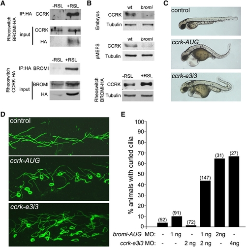
CCRK Interacts with Bromi and Acts in Vertebrate Ciliogenesis
(A) HA-tagged Bromi or HA-tagged CCRK, expressed in NIH 3T3 fibroblasts by Rheoswitch ligand (RSL1)-mediated induction, coimmunoprecipitated endogenous CCRK and Bromi, respectively.
(B) CCRK protein levels in WT versus bromi mutant embryos and embryonic fibroblasts and in uninduced versus RSL1-induced Bromi-HA-expressing NIH 3T3 cells. RSL1 treatment of parental Rheoswitch cells had no effect on CCRK levels (data not shown). Anti-β-tubulin was used as a loading control.
(C) Injection of MOs (4 ng) directed against the start site (ccrk-AUG) and exon 3/intron 3 splice site (ccrk-e3i3) of the zebrafish ccrk homolog resulted in kinking of the body axis and abnormal tail curvature.
(D) Confocal microscopy of acetylated α-tubulin-stained cilia in the posterior (distal early/late) regions of the kidney tubules.
(E) Percentage of animals exhibiting curled cilia. Injection of low doses of bromi-AUG or ccrk-e3i3 MOs (1 ng or 2 ng, respectively) had little effect on their own, whereas coinjection of both MOs at these doses had a synergistic effect. Values in parentheses represent numbers of animals scored.

Acknowledgments
This image is the copyrighted work of the attributed author or publisher, and ZFIN has permission only to display this image to its users. Additional permissions should be obtained from the applicable author or publisher of the image.

Reprinted from Developmental Cell, 18(2), Ko, H.W., Norman, R.X., Tran, J., Fuller, K.P., Fukuda, M., and Eggenschwiler, J.T., Broad-Minded Links Cell Cycle-Related Kinase to Cilia Assembly and Hedgehog Signal Transduction, 237-247, Copyright (2010) with permission from Elsevier. Full text @ Dev. Cell