
- Title
-
Role of Melanocortin Receptor Accessory Proteins in the Function of Zebrafish Melanocortin Receptor Type 2
- Authors
- Agulleiro, M.J., Roy, S., Sánchez, E., Puchol, S., Gallo-Payet, N., and Cerdá-Reverter, J.M.
- Source
- Full text @ Mol. Cell. Endocrinol.
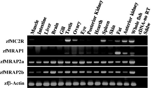
Distribution of zfMC2R and zfMRAPs mRNA expression in different tissues, as revealed by RT-PCR. Ethidium bromide-stained agarose gels showing zebrafish receptor and accessory proteins. Amplifications of β-actin mRNA were used as internal control of the reverse transcription. EXPRESSION / LABELING:
|

(A) Total and cell surface detection of Myc-zfMC2R using anti-c-Myc antibodies. Control corresponds to non-transfected HEK-293/FRT cells. Stable 293/FRT/Myc-zfMC2R cells were transiently transfected with pEGFP (zfMC2R), pcDNA5/FRT/zfMRAP1, pcDNA3/zfMRAP2a, and pcDNA3/zfMRAP2b and assayed for total and extracellular c-Myc detection by whole-cell ELISA. The results represent the mean ± SEM of three independent experiments, each performed in triplicate. Asterisk shows significant differences after one-way ANOVA and Holm–Sidak method (P < 0.05). (B) Immunofluorescence assays in live cells showing the cytoplasmatic expression of zfMC2R in 293/FRT/Myc-zfMC2R cells in absence of MRAPs expression. (C) Immunofluorescence assays in live cells showing the expression of MRAPs (green pseudo-color) and/or zfMC2R (red pseudo-color) in 293/FRT/Myc-zfMC2R cells. Nuclei are counterstained in blue. C- or N-terminally Flag tagged MRAPs were transiently expressed in 293/FRT/Myc-zfMC2R cells. Photomicrographs are taken with a 60x objective and are from a single optical section obtained within an acquisition of z-stacks (0.44 μm/slice) to enable 3D reconstructions of the cells (not shown). Colocalization fluorograms based on 3D reconstructions are shown in the right column. The x-axis of fluorograms is representative of green-labeling and the y-axis is representative of red staining. Colocalization between green-labeling (zfMC2R) and red-labeling (zfMRAPs) voxels are shown as being proportional to each other in fluorograms and correspond to the yellow color in merged slices. |
Reprinted from Molecular and Cellular Endocrinology, 320(1-2), Agulleiro, M.J., Roy, S., Sánchez, E., Puchol, S., Gallo-Payet, N., and Cerdá-Reverter, J.M., Role of Melanocortin Receptor Accessory Proteins in the Function of Zebrafish Melanocortin Receptor Type 2, 145-152, Copyright (2010) with permission from Elsevier. Full text @ Mol. Cell. Endocrinol.