
- Title
-
Nkx6.1 and nkx6.2 regulate alpha- and beta-cell formation in zebrafish by acting on pancreatic endocrine progenitor cells
- Authors
- Binot, A.C., Manfroid, I., Flasse, L., Winandy, M., Motte, P., Martial, J.A., Peers, B., and Voz, M.L.
- Source
- Full text @ Dev. Biol.

Pancreatic expression patterns of nkx6.1 and nkx6.2. Left panel: Whole-mount in situ hybridizations (WISHs) showing expression of nkx6.1 (A,C,E, G) and nkx6.2 (B,D,F, H) at 4 somites (A,B), 18 somites (C,D), 24 hpf (E,F), and 48 hpf (G,H). Right panel: Double fluorescent WISHs showing expression of nkx6.1 and foxa1 (A–C) or neuroD (nrd) (G–I) and of nkx6.2 and foxa1 (D–F) or neuroD (J–L) at 6 somites. Z-plane confocal images. All views are lateral with anterior part to the left. Scale: 20 μm. EXPRESSION / LABELING:
|

Comparison of nkx6.1 and nkx6.2 pancreatic expression. Double fluorescent WISHs showing expression of nkx6.1 and nkx6.2 at 6 somites (A–C), 12 somites (D–F), and 18 somites (G–I). The arrowhead points to a cell expressing both nkx6.1 and nkx6.2 at 12 somites. Z-plane confocal images. All views are lateral with anterior part to the left. Scale: 20 μm. EXPRESSION / LABELING:
|

nkx6.1 and nkx6.2 pancreatic expression at 24 hpf. A–P: Double fluorescent WISHs showing expression at 24 hpf of nkx6.1 and glucagon (gcg) (A), insulin (ins) (B), somatostatin (sst) (C), ghrelin (ghr) (D), neuroD (E), sox4b (F) or pdx1 (G), of nkx6.2 and glucagon (H), insulin (I), somatostatin (J), ghrelin (K), neuroD (L), sox4b (M) or isl1 (N) and of isl1 and sox4b (O) or all the pancreatic hormone genes (P). Z-plane confocal images. Scale: 20 μm. Q: Schematic representation of pancreatic organization at 24 hpf. All views are lateral with anterior part to the left. h: hypochord. EXPRESSION / LABELING:
|

Effects of nkx6 knockdown on endocrine cells. A–P: Ventral views of WISHs showing expression of glucagon at 30 hpf (A, E, I and M), insulin at 24 hpf (B, F, J and N), somatostatin at 24 hpf (C, G, K and O), and ghrelin at 30 hpf (D, H, L and P) in control (A–D), nkx6.1 (E–H), nkx6.2 (I–L), and nkx6.1/nkx6.2 (M–P) morphants. Scale: 20 μm. Q–S: Number of cells expressing glucagon at 30 hpf in nkx6.1 (Q) and nkx6.2 (R) morphants (the horizontal line represents the mean) and number of cells expressing the different pancreatic hormones at 30 hpf in nkx6.1/nkx6.2 morphants (S). The presented data are representative of at least three reproducible and independent experiments. |

Synergistic effects of nkx6 knockdown on alpha-cells. A–D: Ventral views of WISHs showing expression ofglucagon at 30 hpf in control morphants (A) and in morphants injected with 0.3 ng Mo2 6.1 (B), with 0.1 ng Mo1 6.2 (C), or with both simultaneously (D). Scale: 20 μm. E: Number of glucagon-expressing cells in these various morphants (the horizontal line represents the mean). The presented data are representative of at least two reproducible and independent experiments. F: Quantitative analysis of glucagon and insulin expression in 30 hpf embryos, expressed as percentages (100% being arbitrarily fixed as the number of cells in control embryos), following simultaneous injections of Mo2 6.1 and Mo1 6.2 at increasing concentrations. The presented data are representative of at least three reproducible and independent experiments. |

nkx6.1 and nkx6.2 do not regulate each other. A–D: WISHs showing expression of nkx6.2 in control (A) and nkx6.1 (B) morphants, and of nkx6.1 in control (C) and nkx6.2 (D) morphants. E–G: Double fluorescent WISHs showing expression of nkx6.1 and nkx6.2 in control (E), nkx6.1 (F), and nkx6.2 (G) morphants; Z-plane confocal images. The presented data are representative from at least three reproducible and independent experiments. All views are lateral views of 24 hpf embryos with anterior part to the left. Scale: 20 μm. EXPRESSION / LABELING:
|
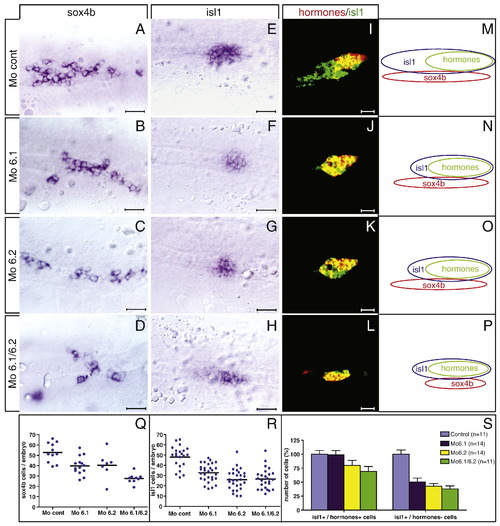
The nkx6 genes are involved in producing/maintaining the endocrine progenitor pool. A–H: WISHs showing expression of sox4b at 18 somites (A–D) and of isl1 at 24 hpf (E–H) in control (A, E), nkx6.1 (B, F), nkx6.2 (C, G), and nkx6.1/nkx6.2 (D, H) morphants. I–L: Double fluorescent WISHs showing expression of isl1 and of all the pancreatic hormone genes together in control (I), nkx6.1 (J), nkx6.2 (K), and nkx6.1/nkx6.2 (L) morphants; Z-plane confocal images. All views are ventral with anterior part to the left. Scale: 20 μm. M–P: Schematic representations (lateral view) of the isl1-, sox4b-, and hormone-expressing cell populations in control and nkx6 morphants. Q: Quantitative analysis of the number of sox4b-expressing cells at 18 somites in control and nkx6 morphants. R: Quantitative analysis of the number of isl1-expressing cells at 24 hpf in control and nkx6 morphants (the horizontal line represents the mean). S: Quantitative analysis of the number of hormone-expressing and hormone-non-expressing isl1-cells at 24 hpf, expressed as percentages (100% being arbitrarily fixed as the number of cells in control embryos), in control and nkx6 morphants. The presented data are representative of at least three reproducible and independent experiments. EXPRESSION / LABELING:
PHENOTYPE:
|

The nkx6 genes are required to the establishment of the endocrine progenitor pool. A–P: WISHs showing expression at 13 somites of pdx1 (A, E, I and M), neuroD (B, F, J and N), sox4b (C, G, K and O), and pax6b (D, H, L and P) in control (A–D), nkx6.1 (E–H), nkx6.2 (I–L), and nkx6.1/nkx6.2 (M–P) morphants. Except D, H, L and P which are lateral, all views are ventral; anterior part to the left. Scale: 20 μm. Q–T: quantitative analysis of the number of pdx1- (Q), neurod- (R), sox4b- (S) and pax6b-expressing cells (T) at 13 somites in control and nkx6 morphants (the horizontal line represents the mean). The presented data were collected from at least two reproducible and independent experiments. In the figure, the superior titles are "Pdx1", "NeuroD", "Sox4b" rather than "Pdx1N", "euroDS" and "ox4b". You will find this figure, at slightly reduced size, at the following address: https://edc.ulg.ac.be/merci/fig8_b5c4cd3b3b244ee41533_.eps. EXPRESSION / LABELING:
PHENOTYPE:
|
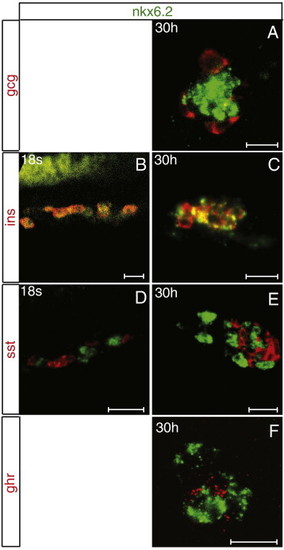
Comparison of nkx6.2 expression with that of pancreatic hormone genes at various stages. Double fluorescent WISHs showing expression of nkx6.2 and glucagon (A), insulin (B–C), somatostatin (D–E), or ghrelin (F) at 18 somites (B, D) and at 30 hpf (A, C, E, F). Z-plane confocal images. A, C–F: Ventral views. B: Lateral views with anterior part to the left. Scale: 20 μm. |

Effects of the nkx6 knockdown on embryo general morphology and on the whole endoderm. A–D: bright-field pictures of control (A) nkx6.1 (B), nkx6.2 (C) and nkx6.1/nkx6.2 (D) morphants at 24 hpf. E–L: WISHs showing expression of foxa1 at 30 hpf (E–H) and myoD at 13 somites (I–L) in control (E; I), nkx6.1 (F; J), nkx6.2 (G; K) and nkx6.1/nkx6.2 (H; L) morphants; all views are ventral with anterior part to the left. Scale: 20 μm. |

neuroD, sox4b and pax6b pancreatic expression at 14 s. Double fluorescent WISHs showing expression at 14 somites of neuroD and sox4b (A–C) or pax6b (D–F). Z-plane confocal images. Scale: 20 μm. All views are lateral with anterior part to the left. |
Reprinted from Developmental Biology, 340(2), Binot, A.C., Manfroid, I., Flasse, L., Winandy, M., Motte, P., Martial, J.A., Peers, B., and Voz, M.L., Nkx6.1 and nkx6.2 regulate alpha- and beta-cell formation in zebrafish by acting on pancreatic endocrine progenitor cells, 397-407, Copyright (2010) with permission from Elsevier. Full text @ Dev. Biol.