
- Title
-
Transgenic Labeling of Hair Cells in the Zebrafish Acousticolateralis System
- Authors
- McDermott, B.M., Asai, Y., Baucom, J.M., Jani, S.D., Castellanos, Y., Gomez, G., McClintock, J.M., and Hudspeth, A.J.
- Source
- Full text @ Gene Expr. Patterns
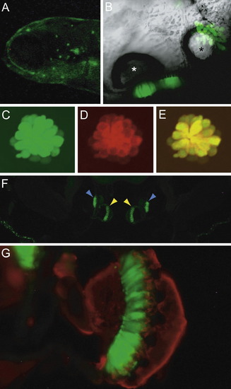
Cellular distribution of GFP in the Ppv3a-3 transgenic line. (A) A fluorescent image of a 5-dpf larval head demonstrates labeling of neuromasts in the anterior lateral line. (B) In a ventral view of an otocyst in a 5-dpf larva, sensory epithelia containing GFP-positive hair cells are located beneath the two otoliths. The anterior otolith is marked with a white asterisk, the posterior otolith with a black one. (C) Transgenically expressed GFP marks hair cells in a 5-dpf larval neuromast. (D) Labeling with FM4-64, a fluorophore that passes through functional mechanotransduction channels, reveals the complement of mature hair cells. (E) The merged image of GFP and FM4-64 signals confirms that the transgenic label marks all the functional hair cells. (F) A transverse section of the head of an adult fish portrays the GFP-positive sensory maculae of the medial sacculi (yellow arrowheads) and the lateral lagenae (blue arrowheads). (G) A higher-magnification image of one sacculus shows GFP-positive hair cells (green) and nerve fibers labeled with an antiserum against neurofilaments (red). EXPRESSION / LABELING:
|

Labeling pattern in a strain generated with the Ppv3b-4 construct. (A) In the Ppv3b-4 line, GFP expression occurs in the anterior macula (AM), posterior macula (PM) and lateral-line neuromasts, of which a representative example is circled. Ectopic peridermal expression is also apparent. (B) At 5 dpf, fluorescence is observed in hair cells of infraorbital neuromast 3 (Raible and Kruse, 2000). (C) In a 5-dpf transgenic larva, the L1 neuromast of the posterior lateral line (Ghysen and Dambly-Chaudiere, 2004) shows much stronger GFP expression in hair cells than in the periderm. (D) The anterior macula of a transgenic fish fixed at 2 dpf and labeled with phalloidin (red) shows GFP expression in the presumptive hair cells prior to hair-bundle formation. The weak phalloidin signal may represent microvilli on the apical surfaces of hair cells and supporting cells. (E) Phalloidin labeling (red) of the anterior macula in a 5-dpf larva reveals the presence of hair bundles (HB) on transgene-labeled hair cells. The scale bars represent 5 μm. |

Transgene expression in larvae of strain Ppv3b-4-12825. (A) The transgenic line Ppv3b-4-12825 displays a pattern of expression similar to that of the Ppv3b-4 line in the ear and lateral line; however, labeling also extends to the eye and gut. (B) In supraorbital neuromast 3 of a 7-dpf embryo, the overlap of GFP expression (green) and immunolabeling for parvalbumin 3 (red) causes hair cells to appear yellow. Some supporting cells (blue arrowheads) and mantle cells (yellow arrowheads) also express GFP. (C) Similar labeling of the posterior macula at 7 dpf reveals GFP expression (green) primarily in the supporting cells separating hair cells labeled for parvalbumin 3 (red). (D) In the crista of a horizontal semicircular canal at 30 dpf, labeling like that above demonstrates robust expression of GFP (green) in supporting cells and of parvalbumin 3 in hair cells (red). Blue and yellow arrowheads designate supporting and mantle cells, respectively. The scale bars represent 5 μm. EXPRESSION / LABELING:
|

GFP expression pattern in a strain generated with the Ppv3b-3 construct. (A) Phalloidin labeling (red) of the anterior macula in a 5-dpf larva shows the presence of a hair bundle on a transgene-labeled hair cell. (B) In a transgenic fish fixed at 2 dpf and labeled with phalloidin (red), GFP expression reveals a presumptive hair cell of the anterior macula prior to hair-bundle formation. The scale bars represent 2 μm. |

The pattern of GFP expression in a line generated with the Ppv3b-3 construct. (A) Phalloidin labeling (red) of the posterior crista in a 5-dpf larva demonstrates a hair bundle on a transgene-labeled hair cell. (B) In the posterior crista of a transgenic fish fixed at 5 dpf and labeled with phalloidin (red), GFP expression reveals a presumptive hair cell prior to hair-bundle formation. The scale bars represent 2 μm. EXPRESSION / LABELING:
|

Unillustrated author statements EXPRESSION / LABELING:
|
Reprinted from Gene expression patterns : GEP, 10(2-3), McDermott, B.M., Asai, Y., Baucom, J.M., Jani, S.D., Castellanos, Y., Gomez, G., McClintock, J.M., and Hudspeth, A.J., Transgenic Labeling of Hair Cells in the Zebrafish Acousticolateralis System, 113-118, Copyright (2010) with permission from Elsevier. Full text @ Gene Expr. Patterns