
- Title
-
Transcriptional regulatory regions of gap43 needed in developing and regenerating retinal ganglion cells
- Authors
- Kusik, B.W., Hammond, D.R., and Udvadia, A.J.
- Source
- Full text @ Dev. Dyn.

The 708-bp promoter is sufficient to promote expression in developing but not regenerating retina. Native GFP expression (green) and DAPI staining (blue) in transverse sections of embryonic, larval, and adult retinas. The GfG43SA and GfG43-708 constructs both express GFP in the retinal ganglion cell layer (gcl) and their axons (*) observed in the fiber layer at 48 hpf (A,E) and in the inner plexiform layer (ipl) within the retina at 96 hpf (B, F). In control and regenerating adult retina (7 days post-crush) of the GfG43SA line, low levels of GFP are observed in control retina (C) and high levels in the GCL and fiber layer of regenerating retina (D). In the GfG43-708 line, there is no GFP expression in the GCL of either control (G) or regenerating (H) retina. Developing retina scale bar (F) = 25 μm. Adult retina scale bar (H) = 50 μm. |

Changes in developmental expression pattern as a result of promoter deletions. Transgene expression in stable GFP reporter fish carrying the Fugu 3.6-kb gap43 promoter (A-D), or promoter deletions: ΔA (E-H), ΔB (I-L), ΔAB (M-P), and ΔABC (Q-T). Images of 48-hpf embryos captured on the fluorescent stereoscope (A, A inset, E, I, M, Q) show that promoter deletions result in overall lower levels of GFP expression (scale bar in Q = 500 μm). The image in (A) was taken at 1/3 the exposure time, while the overexposed image in the inset was taken at the same exposure time as E, I, M, Q. Higher magnification views of the embryos (B-D, F-H, J-L, N-P, R-T; scale bar in T = 100 μm). Dorsal view of hindbrain at 24 hpf (B, F, J, N, R). Lateral view of brain at 48 hpf (C, G, K, O, S). Ventral view of head at 48 hpf (D, H, L, P, T). cb, cerebellum; hb, hindbrain; ob, olfactory bulb; oc, optic chiasm; oe, olfactory epithelium; ot, optic tectum; rgc, retinal ganglion cells. |
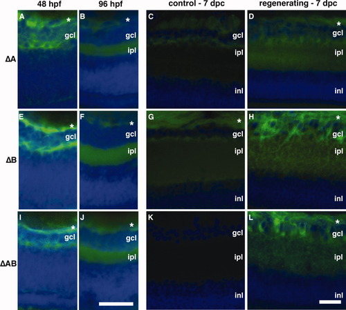
Transgene expression in regenerating retina requires region C. Native GFP expression (green) and DAPI staining (blue) in transverse sections of embryonic (48 hpf; A, E, I), larval (4 dpf; B, F, J), and adult retinas (control; C, G, K; and regenerating; D, H, L) from promoter deletion GFP reporter lines: ΔA (A-D); ΔB (E-H); ΔAB (I-L). All lines displayed the same spatial and temporal pattern of expression in the developing retina (A, B, E, F, I, J) as previously observed for the full-length promoter (see Fig. 2A, B). Unlike ΔABC reporter lines, which did not express GFP in regenerating adult retina (see Fig. 2G, H), addition of the C region in the ΔAB lines was sufficient to restore regenerative expression (L). (*) Axons of RGCs in fiber layer. dpc, days post-crush; gcl, ganglion cell layer; ipl, inner plexiform layer; inl, inner nuclear layer. Developing retina scale bar (J) = 25 μm. Adult retina scale bar (L) = 50 μm. |