
- Title
-
Churchill and Sip1a repress fibroblast growth factor signaling during zebrafish somitogenesis
- Authors
- Kok, F.O., Shepherd, I.T., and Sirotkin, H.I.
- Source
- Full text @ Dev. Dyn.

ChCh and Sip1a are required for somitogenesis. A-S: Living wild-type, and ChCh and Sip1a compromised embryos. A-E: Early epiboly movements appear normal in chch or sip1a morphants. F-J: By 12 somites, misshapen somites are apparent in both chch and sip1 morpholino treated embryos. K-O: In these embryos, somites are less extended through the anterior-posterior axis and overextended through the mediolateral axis. Horizontal and vertical red dotted lines span the width and length of the first four wild-type somites for comparison to the first four somites of morphant embryos, which are marked with a black line. P-S: At 24 hours post fertilization (hpf), somites are enlarged and misshapen in both ChCh and Sip1a compromised embryos. Arrowheads denote notochord and black arrows denote somites. A-J,P-S are lateral views; K-O, dorsal views. PHENOTYPE:
|
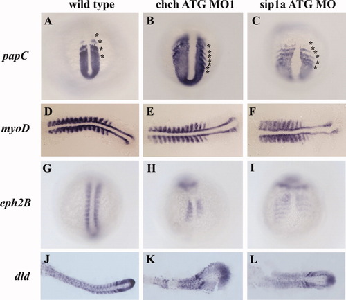
Patterning of the presomitic and somitic mesoderm is disrupted in ChCh and Sip1a compromised embryos. A-L: Whole-mount (A-C,G-I) and flat-mount (D-F,J-L) RNA in situ hybridization of somite markers in wild-type, ChCh and Sip1a compromised embryos. All views are dorsal; anterior to the top (A-C,G-I) and anterior to the left (D-F,J-L). A-C: The expression domains of the PSM marker, papC is broader mediolaterally in ChCh and Sip1a compromised embryos than the wild-type siblings. The number of the papC expression stripes corresponding to the prospective and formed somites at the segmentation plate in chch and sip1a MO is higher than in wild-type siblings (compare asterisks number). D-F: The myogenic regulatory factor, myoD, is expressed in the posterior somite compartment in chch and sip1a MO injected embryos as in wild-type embryos. G-L: ephB2 and dld expression at the anterior half of the somites is reduced and diffuse. Asterisks denote (prospective) somites. EXPRESSION / LABELING:
PHENOTYPE:
|

Inhibition of ChCh and Sip1a affects the components of the "clock and wavefront model." A-I: Flat-mount RNA in situ hybridization of 10-somite stage wild-type, and ChCh and Sip1a compromised embryos. All views are dorsal; anterior to the left. A-F: In wild-type embryos, periodic activation of Notch signaling provides cycling gene expression of Notch pathway genes such as her1 and her7. The number of the her7 expressing stripes in chch and sip1a ATG MO injected embryos ranges from 4 to 5 (B,C; E,F). G-I: fgf8 expression domain at tail bud is expanded anteriorly in ChCh and Sip1a compromised embryos. A-F: Asterisks denote each her1 or her7 expressing premesoderm stripe. G-I: Arrows denote fgf8 tail bud expression domain (G-I). EXPRESSION / LABELING:
PHENOTYPE:
|

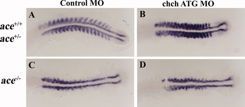
Somite malformation in ChCh and Sip1a compromised embryos can be rescued by reduction of fibroblast growth factor-8 (FGF8). A,B: Dorsal views of 10-somite stage (A) and 12-somite stage (B) living embryos, anterior to the top. A: Somites are narrower at the anterior-posterior (A/P) axis, broader at the mediolateral axis in wild-type and ace heterozygous chch morphants, but not ace homozygous chch morphants. ace homozygous sip1a ATG morphants do not have somite phenotype, but wild-type and ace heterozygous sip1a morphants still have the somite defects. B: Horizontal and vertical red dotted line spans the first five somites of the control injected embryo width and length for comparison to the first five somite of morphant embryos which is indicated with a black line. Embryos were genotyped for the ace allele after imaging. PHENOTYPE:
|
Somite defects in ace mutants are unaltered by ChCh knockdown. A-D: Flat-mount RNA in situ hybridization of myoD in 17-somite stage ChCh compromised wild-type, ace+/-, and ace-/- embryos. All views are dorsal; anterior to the left. C: Lateral myoD expression in the somites is lost in ace mutant embryos. D: myoD expression pattern in ChCh compromised ace-/- embryos is similar to control MO injected ace-/- siblings. EXPRESSION / LABELING:
|
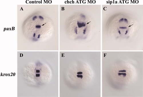
Repression of fibroblast growth factor-8 (FGF8) by ChCh is not limited to the mesoderm. A-F: Whole-mount RNA in situ hybridization of pax2a and krox20 in 10-somite stage ChCh and Sip1a compromised embryos. All views are dorsal; anterior to the top. A-C: FGF target gene pax2a is expanded anteriorly in the isthmus in chch morphants, but not in sip1a morphants. D-F: The anterior extent of krox20 expression is not altered by chch or sip1a knockdown. Arrows denote isthmus. EXPRESSION / LABELING:
|
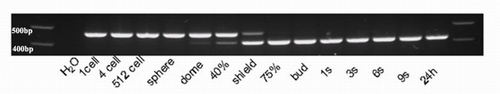
sip1a splice morpholino is effective in eliminating wild-type sip1a mRNA. Reverse transcriptase-polymerase chain reaction (RT-PCR) of mRNA from staged sip1a splice MO injected embryos with sip1a specific primers reveals that the smaller misspliced product is detected by dome stage (4.3 hours postfertilization [hpf]). By 75% epiboly (8 hpf), no wild-type mRNA is detected. |
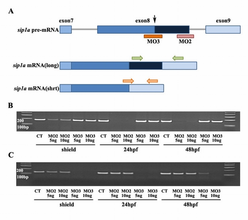
sip1a splice variant targeting morpholinos efficiently eliminate short and long forms. A: Diagram of the alternative splicing within the 3′ region of the sip1a pre-mRNA. Alternative splicing of exon 8 eliminates one zinc finger (dark blue box), which is present in the longer form. Sip1a MO3 (orange bar) targeted to alternative splice site in exon 8 (denoted by black arrow) blocks the alternating splicing event and eliminates production of the short form. Sip1a MO2 (pink bar) blocks the pre-mRNA splicing event needed to generate the long form by targeting the splice site at the 3′ end of exon 8. B,C: Reverse transcriptase-polymerase chain reaction (RT-PCR) of mRNA from staged sip1a splice MO2 and MO3 injected embryos with sip1a full length specific primers (B, green arrows in A) and sip1a short form specific primers (C, orange arrows in A ). B,C: sip1 splice MO2 efficiently altered splicing so that the full length message is eliminated and only the shorter form was produced (B) while sip1 splice MO3 blocked production of the shorter form (C). |
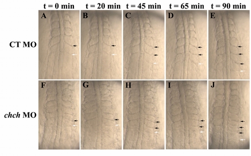
Pace of the "molecular clock" is not significantly altered in ChCh-compromised embryos. Living wild-type and ChCh-compromised embryos beginning at the seven-somite stage. A-J: All views are dorsal, anterior to the top. Duration of somite formation in both control (A-E) and ChCh-compromised embryos (F-J) is approximately 45 min at 23°C. Black arrows denote already formed somite boundaries and white arrows denote newly forming segmentation furrow. |