
- Title
-
WNT5A mutations in patients with autosomal dominant Robinow syndrome
- Authors
- Person, A.D., Beiraghi, S., Sieben, C.M., Hermanson, S., Neumann, A.N., Robu, M.E., Schleiffarth, J.R., Billington, C.J. Jr, van Bokhoven, H., Hoogeboom, J.M., Mazzeu, J.F., Petryk, A., Schimmenti, L.A., Brunner, H.G., Ekker, S.C., and Lohr, J.L.
- Source
- Full text @ Dev. Dyn.
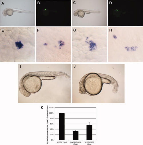
Cysteine mutations in WNT5A reduce the ability of this protein to activate noncanonical Wnt signaling in zebrafish embryos. A,B: Uninjected brightfield (A) and fluorescent images of insulin-enhanced green fluorescent protein (eGFP) transgenic zebrafish embryos (B) at 30 hours postfertilization (hpf) showing insulin-positive cells have coalesced to form an islet. C,D: Insulin-eGFP-positive cells in embryos injected with WNT5A mRNA do not coalesce properly, providing an in vivo assay for noncanonical Wnt5a activity. E: In situ hybridization showing insulin-expressing cells coalescing to form the zebrafish islet at 30 hpf. F-H,K: Injections with WNT5A mRNA (F),WNT5AC182R mRNA (G,H), or WNT5AC83R mRNA (not shown) show scattered insulin-expressing cells at 30 hpf, but the efficacy of this effect is reduced in WNT5AC182R (32%±3% n = 93; P = 0.00005) and WNT5AC83R (55% ± 9% n = 152; P = 0.002) mRNA injected embryos compared with WNT5A mRNA injected embryos (100% ± 6% n = 260) (K). I: Uninjected zebrafish embryo at 26 hpf. J: Zebrafish embryo at 26 hpf injected with dnWNT5A mRNA (25 pg) showing a bent tail phenotype similar to pipetail (Wnt5) mutants. EXPRESSION / LABELING:
|