
- Title
-
Zebrafish zic2a patterns the forebrain through modulation of Hedgehog-activated gene expression
- Authors
- Sanek, N.A., Taylor, A.A., Nyholm, M.K., and Grinblat, Y.
- Source
- Full text @ Development

Hh signaling activates six3b expression in the developing zebrafish forebrain. (A,B) At 15s, six3b is expressed normally in the anterior forebrain of vehicle-treated embryos (black arrowheads in A, 15/15 embryos) and dramatically reduced in cyclopamine-treated embryos (red arrowheads in B, 14/14 embryos treated from the 2- to 4-cell stage onwards). (C,D) At prim-10, ventral forebrain six3b expression is strong in wild-type (WT) embryos (black arrowheads in C, 67/92 embryos) and absent or greatly reduced in smo-/- mutants (red arrowheads in D, 25/92 embryos). (E-H) Embryos injected with a low amount of shha mRNA (12.5 pg) show expanded six3b expression [green arrowheads in H, 10/23 embryos, 2 experiments (n=2)], but no change in ptc1 (F, 31/31 embryos, n=2). Embryos shown in panels A-H are lateral views. Panels A′-H′ are ventral views of the same embryos, anterior to the left. EXPRESSION / LABELING:
PHENOTYPE:
|

Zic2a restricts six3b expression throughout the forebrain beginning at mid-somitogenesis. (A,B) six3b is similarly expressed in conMO- and zic2aMO-injected embryos at 10s-12s (A, 41/41 embryos, n=2; B, 57/57 embryos, n=2). (C,D) At 12s-15s, six3b expression is normal in control morphants (black arrowheads in C, 38/38 embryos, n=3), and expanded throughout the forebrain in zic2a morphants (green arrowheads in D, 58/58 embryos, n=3). (E,F) six3b expansion in zic2aMO-injected embryos persists at prim-5 (black arrowheads in E, 42/42 control morphants, n=3; green arrowheads in F, 40/41 embryos, n=3). (G) Real-time PCR showed that six3b levels, normalized to gapdh, were increased (P=0.0001) in ZicMO-injected embryos relative to control morphants (based on three biological replicates, with two technical replicates per biological replicate). A-F are lateral views; eyes have been removed from embryos in C-F. A′-B′ are ventral views of the same embryos. C′-F′ are dorsal views of the same embryos, anterior to the left. EXPRESSION / LABELING:
PHENOTYPE:
|
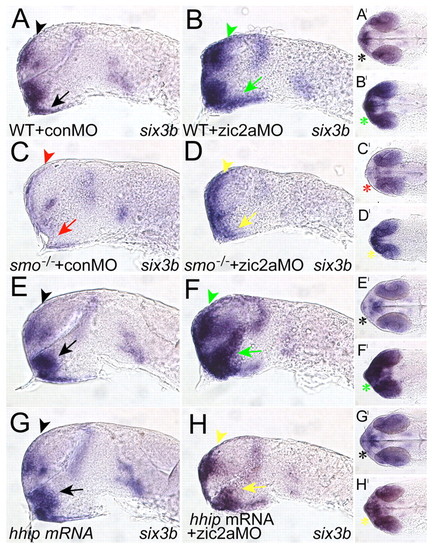
Reduced Hh signaling in zic2a morphants prevents six3b expansion. (A-D) Zic2a depletion in smo-/- mutant embryos. Zic2a-depleted WT siblings show expanded six3b in the telencephalon and diencephalon (green arrowheads in B, 120/151 embryos, n=2), whereas smo-/- mutants injected with conMO lose six3b expression (C, 25/25 smo-/- embryos, n=2). zic2aMO-injected smo-/- mutants show upregulated, but anteriorly restricted, six3b expression (yellow arrowheads in D, 14/17 smo-/- embryos, n=2). (E-H) Zic2a depletion in the presence of exogenous Hhip. Embryos injected with zic2aMO alone show expanded six3b (green arrowheads in F, 45/45 embryos, n=2), whereas embryos injected with hhip mRNA express six3b normally (black arrowheads in G, 43/43 embryos, n=2). Embryos co-injected with zic2aMO and hhip mRNA show rescued six3b expression (yellow arrowheads in H, 17/40 embryos, n=2). A-H are lateral views, anterior to the left. A′-H′ are ventral views of the same embryos, anterior to the left. Arrowheads point to the telencephalon and arrows to the diencephalon. Asterisks in A′-H′ illustrate six3b expression in the optic stalks. All embryos are shown at prim-5. |

Six3b depletion rescues prethalamic patterning defects in Zic2a-depleted embryos. (A) Uninjected embryos show normal dlx2a expression in the diencephalon (29/29 embryos, n=2). (B) zic2a morphants exhibit a strong reduction of diencephlic dlx2a (18/19 embryos, n=2). (C) six3b morphants show normal dlx2a patterning (23/23 embryos, n=2). (D) Embryos co-injected with six3bMO and zic2aMO exhibit partially rescued prethalamic dlx2a expression (30/52 embryos, n=2). (E) A graphic summary of the dlx2a rescue experiments. (F) Normal arx expression is observed in the PT of uninjected control embryos. (G) arx expression is strongly reduced in zic2a morphants (13/15 embryos, n=2). (H) six3b morphants show normal arx expression. (I) Embryos co-injected with six3bMO and zic2aMO show partial rescue of arx (10/23 embryos, n=2). (J) A graphic summary of the arx domain rescue experiments. Red, strongly reduced dlx2a or arx in PT; blue, normal or nearly normal expression of dlx2a or arx in PT. Arrowheads point to the diencephalic domains of dlx2a or arx expression. Lateral views with anterior to the left shown at prim-5 (A-D) or 20s (F-I). EXPRESSION / LABELING:
|

Zic2a limits distal OS marker expression in the ventral retina. The effect of Zic2a depletion was examined using ISH. (A-D) The following markers were expressed normally in control and zic2a morphants: rx3 in the pre-optic area of the hypothalamus (A, 36/36 control morphants, n=3; B, 42/44 zic2a morphants, n=3); vax1 in the proximal OS (C, 50/50 control morphants, n=4; D, 58/68 zic2a morphants, n=4). (E,F) pax2a is expressed normally in control morphants (E, 51/52 embryos, n=4), and is expanded into the retina in zic2a morphants (F, 123/131 embryos, n=6). (G,H) fgf8a is restricted to the OS in control morphants (G, 20/20 embryos, n=2), and expanded into the retina when Zic2a is depleted (H, 44/53 embryos, n=3). Green arrowheads in F,H point to abnormal expression in the ventral retina. A-H are lateral views, anterior to the left. A′-H′ are ventral views of the same embryos, anterior to the left. All embryos are at prim-5. EXPRESSION / LABELING:
|

Ventral retinal defects in zic2a morphants. (A,B) pax2a is normally expressed at the OS-retinal border of control morphants (A) and is expanded into the ventral retina of zic2a morphants (B). (C,D) pax6a expression is seen throughout the retina of Tg(-8.0cldnb:lynGFP)zf106 embryos injected with conMO (C) or zic2aMO (D). Arrowheads in C,D point to the anterior limit of pax6a expression, and asterisks mark the posterior limit of cldnb:gfp expression in the nasal retina. (E,F) aldh1a3 expression is normal in conMO-injected embryos (arrowheads in E, 44/44 embryos, n=2). The zic2a morphant retina fails to close and aldh1a3 expression is expanded (arrowheads in F, 35/38 embryos, n=2). The choroid fissure is closed in uninjected embryos (arrowhead in G), but open in zic2a morphants (red arrowheads in H, 21/28 embryos, n=2). A-F show dissected retinae at prim-5, anterior to the left. A-D are confocal z-stacks. Embryos in G,H are at the high-pec stage |
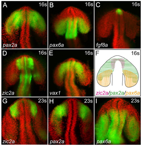
Patterned gene expression in OS and retinal precursors. (A-F) Wild-type embryos at 16s stained using wholemount in situ hybridization (WISH, green) for expression of pax2a (A), pax6a (B), fgf8a (C), zic2a (D) and vax1 (E). Nuclei were counterstained using DAPI (red). The schematic (F) illustrates the overlap between zic2a and pax2a expression domains in the presumptive OS, and the overlap between pax2a and pax6a domains in the presumptive retina. (G-I) Expression of zic2a (G), pax2a (H) and pax6a (I) detected at 23s. Expression patterns, imaged using confocal microscopy, are shown as z-stacks in ventral view, anterior up. EXPRESSION / LABELING:
|

Hh is necessary and sufficient for zic2a expression in the OS and ventral retina. (A-F) zic2a is expressed in the OS at 16s (A). In smo-/- mutants, zic2a expression is reduced in the OS at this stage (B, 14/14 embryos, n=2), but not elsewhere. shha mRNA-injected WT embryos express zic2a ectopically in the OS and retina at 16s (C, 13/13 embryos, n=1). zic2a expression is still reduced in smo-/- mutants at 19s (D,E, 30/30 embryos, n=2), and expanded in shha-injected retina (F, 19/19 embryos, n=1). The same result was observed at prim-5 (39/42 embryos, n=2, data not shown). Arrowheads point to the OS. Embryos are in lateral view, anterior to the left. EXPRESSION / LABELING:
|

Zic2a patterns the OS and retina in an Hh-dependent manner. (A-D) Zic2a-depleted WT siblings show expanded pax2a (B, 99/99 embryos, n=4), whereas smo-/- mutants injected with conMO lose pax2a expression (C, 12/12 embryos, n=2). zic2aMO-injected smo-/- mutants show a rescued pax2a phenotype (D, 30/30 embryos, n=4). (E-H) Co-injection of conMO and shha mRNA, or injection of zic2aMO alone, causes moderately expanded pax2a expression (F, 20/34 embryos, n=2; G, 30/30 embryos, n=2, respectively). Embryos injected with zic2aMO and shha mRNA show strong pax2a expansion (H, 23/33 embryos, n=2). Arrowheads mark the OS. Panels A-H are lateral view, anterior to the left. Panels A′-H′ are ventral views of the same embryos, anterior to the left. All embryos are at prim-5. EXPRESSION / LABELING:
|

Exogenous Hh antagonist rescues OS and retinal patterning in Zic2a-depleted embryos. (A) Wild-type pax2a expression. (B-D) Embryos injected with zic2aMO show expanded pax2a (B, 43/43 embryos, n=3), whereas embryos injected with hhip mRNA show normal pax2a expression (C, 8/8 embryos, n=1). Embryos co-injected with zic2aMO and hhip mRNA show normal pax2a expression (D, 30/41 embryos, n=3). A-D are lateral views, anterior to the left. A′-D′ are ventral views of the same embryos, anterior to the left. Arrowheads mark the posterior limit of pax2a expression in the retina. All embryos are at prim-5. |

zic2a expression in the anterior CNS from 3s-12s. zic2a expression in the OS is first detectable at 3s-4s (A) and continues at 5s-6s (B) and 7s-8s (C) It is not detectable in the adjacent retina at these stages (A-C). Beginning at 11s-12s, zic2a is expressed both in the OS and the distal (dorsal) retina (D, asterisk). A-D are lateral views, anterior to the left. A′-D′ are dorsal views of the same embryos, anterior to the left. |

Apoptosis does not contribute to OS patterning defects observed in Zic2a-depleted embryos. p53MO-injected embryos exhibit normal pax2a expression in the OS (A, 20/20 embryos, n=2). zic2a morphants exhibit strong pax2a expansion into the ventral retina (B, 20/23 embryos, n=2). Embryos co-injected with p53MO and zic2aMO show a similarly strong expansion of pax2a into the adjacent ventral retina (C, 21/21 embryos, n=2). A-C are lateral views, anterior to the left. A′-C′ are ventral views of the same embryos, anterior to the left. All embryos are at prim-5. |

pax2a patterning defects are detected prior to fgf patterning defects in zic2a morphants. fgf8a expression is normal at 16s in control (A, 11/11 embryos) and zic2a morphants (B, 8/8 embryos). Similarly, spry4 is unaffected at 16s in control (C, 21/21 embryos, n=2) or zic2a morphants (D, 22/24 embryos, n=2). pax2a is expressed broadly in the OS and retinal primoridia at 16s (E, 15/15 embryos, n=2), and might be slightly expanded in zic2a morphants (F, 9/21 embryos, n=2). At 19s, fgf8a is weakly expressed in the presumptive OS (G, 12/12 embryos), and its expression is unaffected by knocking-down Zic2a (H, 9/9 embryos). spry4 is expressed in a broader domain than fgf8a (I, 20/20 embryos, n=2), and also appears unaffected in zic2a morphants (J, 21/25 embryos, n=2). pax2a expression has begun retracting from the retinal primordium by 19s in control embryos (K, 8/18, n=2), but not in zic2aMO-injected embryos (L, 9/15 embryos, n=2). Arrowheads mark the OS. All embryos are shown in ventral view, anterior to the left. Embryos in A-F are at 16s, while embryos in G-L are at 19s. |

zic2a acts upstream of Fgf signaling and pax2a in OS development. (A-C) The expression of spry4, a direct target of Fgf signaling, after a 120-minute treatment with vehicle (A, normal in 13/13 embryos, n=2), a 30-minute treatment with SU5402 (B, strongly reduced in 10/10 embryos, n=2) and a 60-minute treatment with SU5402 (C, undetectable in 9/10 embryos, n=2). (D-F) zic2a expression after vehicle treatment for 120 minutes (D, normal in 16/16 embryos), after 30 minutes of SU5402 exposure (E, normal in 19/19 embryos) and after 60 minutes of SU5402 exposure (F, strongly reduced in the telencephalon, 2/17 embryos, but unaffected in the OS, 17/17 embryos). (G,H) zic2a expression at prim-5 after vehicle treatment starting at the tail bud (G, 25/25 embryos), or SU5402 treatment starting at the tail bud (H, absent in the telencephalon, 20/20 embryos, but normal in the OS). (I,J) zic2a is strongly expressed in the OS of both the wild-type (WT; I, 97/97 embryos, n=2) and in pax2a-/- mutant siblings (J, 30/30 embryos, n=2). Arrows point to the telencephalon, arrowheads to the OS. Views are lateral, anterior to the left, except G′ and H′ that are ventral views, anterior to the left, of the embryos pictured in G and H. Embryos are shown at 16s (A-C), 19s (D-F) and prim-5 (G-H). |
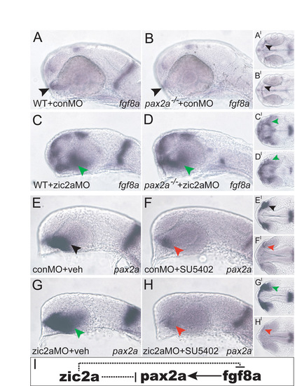
The epistatic relationships between zic2a, pax2a and fgf8a. (A-H) conMO-injected WT siblings exhibit normal fgf8a expression in the OS and MHB (A, 58/60 embryos, n=2). pax2-/- mutant embryos injected with conMO express fgf8a in the OS, but not in the MHB (B, 20/20 embryos, n=2). WT siblings injected with zic2aMO show expanded fgf8a (C, green arrow, 55/80 embryos, n=2). pax2-/- mutants injected with zic2aMO also have expanded fgf8a in the ventral retina (D, 13/23 embryos, n=2), but lack MHB expression. Vehicle-treated embryos injected with conMO display normal pax2a expression (E, 35/36 embryos, n=4), whereas conMO-injected embryos treated with SU5402 show reduced pax2a (F, 34/37 embryos, n=4). Vehicle-treated zic2aMO-injected embryos have expanded pax2a expression (G, 28/32 embryos, n=4), but zic2aMO-injected and SU5402-treated embryos have reduced pax2a expression (H, 33/36 embryos, n=4). (I) The proposed regulatory relationship between pax2a and fgf8a downstream of zic2a. Arrowheads point to the OS. A-H are lateral views, anterior to the left. A′-H′ are ventral views of the same embryos, anterior to the left. Embryos in A-D are at prim-5 and embryos in E-H are at -prim-1. |

Gross retinal patterning and retinal neurogenesis appear unaffected in Zic2a knockdown embryos. (A-H) foxg1 expression in the anterior retina in conMO (A, 20/20 embryos) and Zic2a-depleted embryos (B, 21/21 embryos). efna5a expression in the anterior retina in conMO (C, 32/32 embryos, n=2) and zic2a morphants (D, 30/34 embryos, n=2). atoh7 expression marks the ventral retina in conMO-injected embryos (E, 40/46 embryos, n=2). Expression of atoh7 is absent in zic2aMO-injected embryos (F, red arrow, 49/68 embryos, n=2). Tg(pou4f3:gap43-GFP)s356t embryos allow direct visualization of retinal ganglion cells (RGCs) and their axons in living embryos. RGC differentiation and axon guidance (white arrows) are largely normal in control (G) and Zic2a-depleted embryos (H, 7/7 embryos, n=2). A-D show embryos in dorsal view, anterior at the top. E-F show dissected retina at prim-5, anterior to the left. G and H depict live transgenic embryos at 5 dpf imaged by confocal microscopy, ventral view, anterior up. |
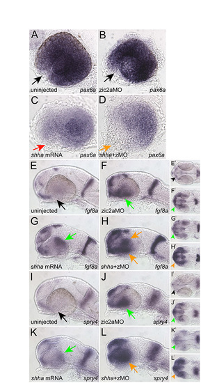
The combined effects of Zic2a depletion and Hh pathway hyperactivation on pax6a, fgf8a and spry4 expression. (A-L) Zic2a knockdown does not affect pax6a expression in the anterior retina (B, normal in 10/10 embryos). By contrast, overexpression of shha inhibits pax6a expression throughout the retina, most noticeably in the nasal retina (C, red arrow, 9/13 embryos). Embryos co-injected with shha mRNA and zic2aMO exhibit an overall reduction of pax6a in the retina, with a more pronounced clearing in the nasal retina (D, orange arrow, 12/12 embryos). fgf8a expression is expanded in zic2aMO-injected embryos (F, green arrow, 11/12 embryos). Similarly, ectopic fgf8a expression is observed in shha-mRNA-injected embryos (G, green arrow, 3/11 embryos). In shha mRNA plus zic2aMO injected embryos, dramatically increased ectopic fgf8a expression is observed (H, orange arrows, 4/12 embryos). spry4 expression is also expanded in zic2aMO-injected embryos (J, green arrow, 10/10 embryos). shha mRNA induces ectopic spry4 (K, green arrow, 11/13 embryos). Co-injection of shha mRNA and zic2aMO leads to a stronger expansion of spry4 expression than either single injection (L, orange arrow, 7/11 embryos). A-D are lateral views of isolated retina, anterior forward. E-L are lateral view, anterior to the left. E′-L′ are ventral views of the same embryos, anterior to the left. All embryos shown at prim-5. |

Six3b depletion does not rescue the retinal patterning defect in Zic2a-depleted embryos. (A) Uninjected embryos show normal pax2a expression in the OS and ventral retina (33/33 embryos, n=2). (B) zic2a morphants exhibit ectopic expression of pax2a in the ventral retina (15/18 embryos, n=2). (C) six3b morphants show normal pax2a patterning (25/25 embryos, n=2). (D) Embryos co-injected with six3bMO and zic2aMO show ectopic pax2a expression in the retina similar to that observed in zic2a morphants (38/46 embryos, n=2). (E) A graphic summary of the pax2a rescue experiments. Red, expanded pax2a; blue, normal pax2a. Arrowheads point to the posterior limit of retinal pax2a expression. Lateral views with anterior to the left shown at prim-5. |