
- Title
-
Abcb10 physically interacts with mitoferrin-1 (Slc25a37) to enhance its stability and function in the erythroid mitochondria
- Authors
- Chen, W., Paradkar, P.N., Li, L., Pierce, E.L., Langer, N.B., Takahashi-Makise, N., Hyde, B.B., Shirihai, O.S., Ward, D.M., Kaplan, J., and Paw, B.H.
- Source
- Full text @ Proc. Natl. Acad. Sci. USA
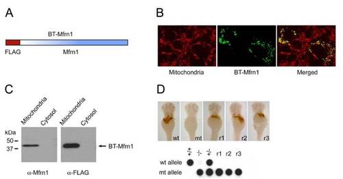
BT-Mfrn1 properly targets to the mitochondrial compartment and complements anemia of frs mutants, and so it is validated as a functional bait for affinity purification. (A) Mouse Mfrn1 was FLAG-tagged at its N terminus as bait for affinity purification. The FLAG-tagged construct was referred as BT-Mfrn1. (B) Immunolocalization of BT-Mfrn1 protein to mitochondria of transfected COS7 cells. Fluorescence confocal images were obtained from immunostained resident mitochondrial proteins (red) and BT-Mfrn1 (green). Colocalized expression of BT-Mfrn1 in the mitochondria is indicated by the yellow signal. (C) BT-Mfrn1 proteins properly targeted to the mitochondrial fraction. Mitochondrial and cytosolic fractions were isolated from BT-Mfrn1-transfected COS7 cells and immunoblotted by anti-Mfrn1 and anti-FLAG antibodies, respectively. (D) Expression of BT-Mfrn1 cRNA rescued the anemia of frascati ( frstq223) embryos. Control wild-type (wt), frs mutant (mt), and rescued frs embryos (r1–3) were stained with o-dianisidine to detect hemoglobinized cells. Control wild-type, mutant, heterozygote, and rescued frs (r1–3) embryos were genotyped. Genotyping results in the Right Lower confirmed that these three putative rescued embryos (r1–3) are mutants. |