
- Title
-
Expression of protocadherin-9 and protocadherin-17 in the nervous system of the embryonic zebrafish
- Authors
- Liu, Q., Chen, Y., Pan, J.J., and Murakami, T.
- Source
- Full text @ Gene Expr. Patterns
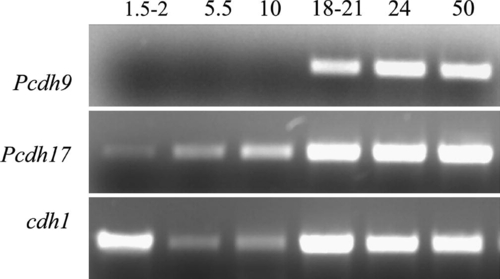
RT-PCR analysis of Pcdh9 and Pcdh17 expression in embryonic zebrafish using total RNAs. RT-PCR for cdh1 was performed as loading control. Numbers represent hours post fertilization. |

Pcdh9 and Pcdh17 expression in 24 hpf zebrafish embryos. All panels show whole mount embryos labeled with Pcdh9 cRNA (A–D, I–L) or Pcdh17 cRNA probes (E–H, M–P). (A and E) Lateral views of the anterior half of the embryos (anterior toward the lower left corner, and dorsal to the upper left corner). (B, C, F and G) Lateral views (anterior to the left and dorsal up) of the head, with (B and F) the fore- and midbrains, and (C and G) showing the hindbrain region. (D and H) Lateral views (anterior to the left and dorsal up) of whole mount eyes. (I, J and M) Frontal views (dorsal up) of embryo heads with focusing planes at the diencephalon level. (K and O) Dorsal views of the hindbrain region (anterior to the left). (L, N and P) Lateral views from the mid-trunk region (anterior to the left and dorsal up). The arrowheads in (B, F, I and M) point to the more anteroventrally located Pcdh9 or Pcdh17 expressing cluster, while the arrows in (B and J) point to the more posterodorsally located Pcdh9 expressing cluster in the anteroventral diencephalon. Asterisks in (C and K) indicate clusters of Pcdh9 expressing cells in the hindbrain. Arrows in (G, H, N, O and P) point to Pcdh17 expressing cells. Abbreviations: c, cerebellum; di, diencephalon; ey, eye; h, hindbrain; hd, head; le, lens; nc, notochord; or, optic recess; ot, optic tectum; ov, otic vesicle; p, pineal organ; rpe, retinal pigmented epithelium; sag, statoacoustic ganglion; sc, spinal cord; te, telencephalon; teg, tegmentum, y, yolk. EXPRESSION / LABELING:
|

Pcdh9 and Pcdh17 expression in 34 hpf embryos. All panels are from whole mount embryos labeled with Pcdh9 cRNA (A–D, I–L) or Pcdh17 cRNA (E–H, M–P) probes. (A and E) Lateral views (anterior to the left and dorsal up) of the anterior 2/3 of the embryos. (B and F) Lateral views (anterior to the left and dorsal up) of the fore- and midbrains, while (C and G) show lateral views of the hindbrain (C shows slightly more anterior structures). (D, H, L and P) Dorsal views (anterior to the left), with (D, H and L) showing mainly the hindbrain, while (P) showing mainly the spinal cord. (I and M) Frontal views (dorsal up) focusing at the telencephalon level, while (J and N) are frontal views (dorsal up) focusing at the diencephalon and tegmentum levels, respectively. (K and O) Lateral views (anterior to the left and dorsal up) of the retina. The asterisk in (B and F) indicates the pretectal area. Segmental expression of Pcdh9 in the hindbrain is indicated by asterisks in (C and D). Abbreviations: ht, hypothalamus; op, olfactory placode. Other abbreviations are the same as in Fig. 4. EXPRESSION / LABELING:
|
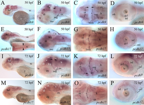
Pcdh9 and Pcdh17 expression in 50 hpf and 72 hpf embryos. (A–D, I–L) From whole mount embryos processed for Pcdh9 in situ hybridization, while (E–H, M–P) are whole mount embryos labeled with Pcdh17 cRNA probe. All lateral views have anterior to the left and dorsal up, while dorsal views have anterior to the left. (A, E, I and M) Lateral views of the anterior half of the embryos showing overall patterns of Pcdh9 or Pcdh17 expression. (B, F, J and N) Lateral views of the head region. (C, G, K and O) Dorsal views of the mid- and hindbrains. (D, H, L and P) Lateral views of the retina. The apposing arrows in (B, C, J and K) indicate the area between the posterior border of the optic tectum and boundary of the mid-hindbrains with reduced Pcdh9 expression. The opposing arrowheads in (E) insert indicate Pcdh17 expression in the spinal cord. Abbreviations: bmh, boundary of mid- and hindbrains; gcl, retinal ganglion cell layer; inl, inner nuclear layer; onl, outer nuclear layer. Other abbreviations are the same as in Fig. 4 and Fig. 5. EXPRESSION / LABELING:
|
Reprinted from Gene expression patterns : GEP, 9(7), Liu, Q., Chen, Y., Pan, J.J., and Murakami, T., Expression of protocadherin-9 and protocadherin-17 in the nervous system of the embryonic zebrafish, 490-496, Copyright (2009) with permission from Elsevier. Full text @ Gene Expr. Patterns