
- Title
-
Expression of five frizzleds during zebrafish craniofacial development
- Authors
- Sisson, B.E., and Topczewski, J.
- Source
- Full text @ Gene Expr. Patterns
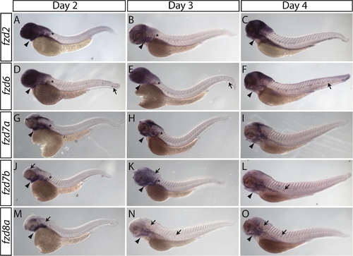
Lateral views of zebrafish fzd2, 6, 7a, 7b, and 8a mRNA expression at 2, 3, and 4 days post-fertilization (dpf). (A–C) fzd2 is expressed within the pharyngeal arches (arrowheads), throughout the head and within the pectoral fin bud (asterisks in A,B). (D–F) fzd6 is expressed within the pharyngeal arches (arrowheads), throughout the head, within the posterior lateral line (arrows in D–F) and within the pectoral fin bud (asterisks in D–E). (G–I) fzd7a is expressed within the pharyngeal arches (arrowheads), in the brain and within the pectoral fin bud (asterisks in G–H). (J–L) fzd7b is expressed within the pharyngeal arches (arrowheads), in the anterior and posterior lateral line (arrows in J–L) and within the pectoral fin bud (asterisks in J–K). (M–O) fzd8a is expressed within the pharyngeal arches (arrowheads) and in the anterior and posterior lateral line (arrows in M–O). (M) At 2 dpf fzd8a is also expressed within the midbrain. |
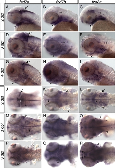
Higher magnification of the gene expression of zebrafish fzd7a, 7b and 8a reveals specific patterning within the head. (A–I) Lateral views of the embryo show all three genes expressed within the pharyngeal arches (arrowheads). (A–C) Gene expression of fzd7a, 7b and 8a at 2 days post-fertilization (dpf). (A) fzd7a is expressed in the dorsal part of the hindbrain (arrow) and within the forebrain (asterisk). (B,C) fzd7b and 8a are expressed within the anterior lateral line (asterisks). (D–F) Gene expression of fzd7a, 7b and 8a at 3 dpf. (D) fzd7a is clearly expressed in the tectum (arrow). (E,F) fzd7b and 8a are expressed within the anterior lateral line (asterisks). (G–I) Gene expression of fzd7a, 7b and 8a at 4 dpf. (G) The gene expression of fzd7a in the dorsal hindbrain is still visible. (J–L) Dorsal views of fzd7a, 7b and 8a gene expression at 2 dpf. (J) A superficial dorsal view of 2 dpf clearly shows the tectum (arrow), rhombic lip (asterisk) and dorsal rhombomere expression (arrowhead) of fzd7a. fzd7b and 8a are expressed at the midline (arrowhead) and in the otic vesicle (arrow) (K and L, respectively). (M–O) Dorsal views of the gene expression of fzd7a, 7b and 8a at 3 dpf. (M) The expression in the in the rhombic lip and dorsal rhombomeres (arrowhead) has moved laterally. (O) The dorsal view of fzd8a expression clearly shows the anterior lateral line expression (asterisks). (P–R) Ventral views of fzd7a, 7b and 8a gene expression at 3 dpf. (P) fzd7a is expressed in the retina adjacent to the lens (arrow). (Q–R) Ventral views of fzd7a and 7b expression reveal expression in the anterior lateral line (asterisk). (R) At 3 dpf fzd8a is expressed within the olfactory bulbs (arrows). Bar equals 100 μm. |
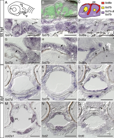
Sections of zebrafish craniofacial cartilage reveal tissue specific gene expression of fzd7a, 7b, and 8a. (A) Schematic of a lateral view of a zebrafish at 55 h post-fertilization (hpf) highlighting the location of pharyngeal arches. Anterior is to the left. (B) Sagittal section at 55 hpf of Tg(fli1a:EGFP)y1/+ that expresses GFP within the neural crest tissue within the pharyngeal arches. Overlay of Nomarski image and fli1a:EGFP expression. (C) Schematic drawing of two pharyngeal arches at 55 hpf illustrating the different gene expression patterns of fzd7a, 7b and 8a. (D–F) Sagittal sections of the pharyngeal arches at 55 hpf. (D) fzd7a is expressed within the neural crest tissue of the pharyngeal arches. (E) fzd7b is expressed within the mesodermal core and the neural crest tissue. (F) fzd8a is expressed within the pharyngeal endoderm of the pharyngeal arches and is excluded from the neural crest and the mesodermal core. Note the similarity between the expression pattern of fzd7a at 55 hpf (D) and the flig1a:EGFP (B). (G–I) At 55 hpf fzd7b, but not fzd7a or 8a, are expressed within the symplectic cartilage elements (arrows). (J–L) At 3 days post-fertilization (dpf) fzd7a, 7b and 8a are no longer expressed within the craniofacial cartilage, but are expressed in the tissue surrounding the cartilage elements. (M) At 3 dpf it is clear that col2a1a is not expressed in the trabeculae but is expressed in the more posterior cartilage elements of the palaquadrate and Meckel’s cartilage. (N–O) Much like fzd7a, 7b and fzd8a, 2 (N) and fzd6 (O) are expressed in the tissue surrounding the cartilage elements and not directly in the cartilage at 3 dpf. Abbreviations: (e) eye, (mc) Meckel’s cartilage, (pq) palaquadrate, (t) trabeculae, (ep) ethmoid plate, (nc) neural crest, (pe) pharyngeal endoderm, (mes) mesodermal core, (ov) otic vesicle, and (p) pharyngeal arches. Bar equals 50 μm. EXPRESSION / LABELING:
|
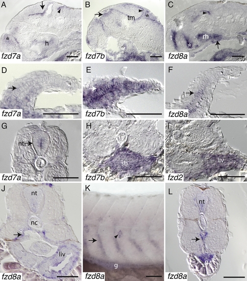
Sagittal sections of 55 h post-fertilization (hpf) reveal specific expression of fzd7a, 7b, and 8a within the brain. (A) fzd7a is expressed within the dorsal hindbrain (arrowhead), forebrain (asterisk), midbrain tegmentum (arrow) and the hypothalamus. (B) fzd7b is expressed within the tectum (arrowhead), rhombic lip (asterisk), thalamus (arrow) and the tegmentum. (C) fzd8a is expressed in the telencephalon, rostal hypothalamus, along the border between the hypothalamus and the ventral thalamus (asterisk), the midbrain tegmentum (arrowhead), and the hyophysis (pituitary) (arrow). (D-F) Transverse sections of the pectoral fin bud at 55 hpf. (D) fzd7a is expressed throughout the pectoral fin bud at a low level (arrow). (E) fzd7b is expressed throughout the pectoral fin bud including the cartilage (arrow). (F) fzd8a is expressed within the cartilage of the pectoral fin bud (arrow). (G) A coronal section of a 55 hpf embryo reveals that fzd7a is expressed within the neural tube (arrow). (H–J) Coronal sections at 55 hpf (H) and 3 dpf (I–J) reveal staining within the gut. (H) fzd7b is expressed in the swim bladder and some mesenchymal tissue surrounding the gut. (I) fzd2 is expressed in the liver. (J) A more posterior coronal section demonstrates that fzd8a is expressed in the liver and along the ventral borders of the myotomes (arrow). (K) A lateral view at 3 dpf illustrates that the expression of fzd8a along the ventral borders of the myotomes is also within the anterior side of each segment of the trunk (arrow). This view also shows the expression of fzd8a within the lateral line (arrowhead) and within the gut. (L) Coronal section at 3 dpf shows expression of fzd8a in the neural tube, gut and in the vasculature (arrow). Abbreviations: (nt) neural tube, (g) gut, (nc) notochord, (liv) liver, (sb) swim bladder, (h) hypothalamus, (tm) tegmentum, (t) telencephalon, and (rh) rostal hypothalamus. Bar equals 50 μm. EXPRESSION / LABELING:
|
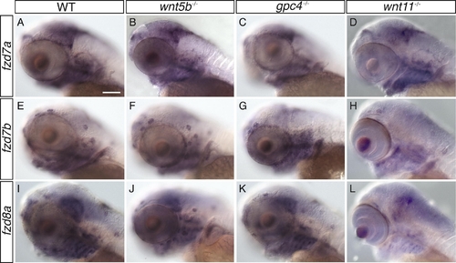
Expression of fzd7a, 7b and 8a within wnt5b, gpc4, and wnt11 mutants is normal at 3 days post-fertilization. (A, E, and I) wild type siblings. (B, F, and J) wnt5b (pipe tail) mutants. (C, G, and K) gpc4 (knypek) mutants (D, H, and L) wnt11 (silberblick) mutants. The different shapes of the wnt11 (silberblick) mutant heads are due to the synophthalmia phenotype. Bar equals 100 μm. EXPRESSION / LABELING:
PHENOTYPE:
|
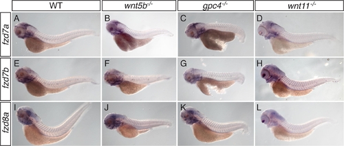
Lateral view of whole embryos illustrates that the expression of fzd7a, 7b and 8a within wnt5b, gpc4, and wnt11 mutants at 3 days post-fertilization is normal. (A, E, and I) wild type siblings. (B, F, and J) wnt5b (pipe tail) mutants. (C, G, and K) gpc4 (knypek) mutants. (D, H, and L) wnt11 (silberblick) mutants. The different shapes of the wnt11 (silberblick) mutant heads are due to the synophthalmia phenotype. PHENOTYPE:
|

Unillustrated author statements EXPRESSION / LABELING:
|
Reprinted from Gene expression patterns : GEP, 9(7), Sisson, B.E., and Topczewski, J., Expression of five frizzleds during zebrafish craniofacial development, 520-527, Copyright (2009) with permission from Elsevier. Full text @ Gene Expr. Patterns