
- Title
-
Identification of vasculature-specific genes by microarray analysis of etsrp/etv2 overexpressing zebrafish embryos
- Authors
- Wong, K.S., Proulx, K., Rost, M.S., and Sumanas, S.
- Source
- Full text @ Dev. Dyn.
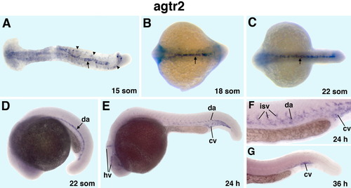
Zebrafish angiotensin II type 2 receptor (agtr2) is expressed in vascular endothelial cells during early embryonic development as analyzed by in situ hybridization. A-C: Dorsal view, anterior to the left. D-G: Lateral view, anterior to the left. A-G: A flat-mounted 15-somite embryo (A), 18-somite (B), 22-somite (C,D), 24 hours postfertilization (hpf; E,F); 36 hpf stage whole-mount embryos (G). F,G: Enlarged views of the tail region. Note agtr2 expression in bilateral endothelial cell precursors (arrowheads, A) and in the axial vessels (arrows, A-C). D: Agtr2 expression is limited mostly to the dorsal aorta (da). E,F: Agtr2 is expressed in the dorsal aorta, head (hv), and intersegmental vessels (isv) and the cardinal vein (cv). G: Agtr2 expression is mostly confined to the cardinal vein plexus. EXPRESSION / LABELING:
|
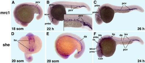
Vascular-endothelial expression of the zebrafish mannose receptor C1 (mrc1) and src homology 2 domain containing E (she) as analyzed by in situ hybridization. A-C,E,F: Lateral view, anterior to the left. D: Anterior view. A-C: mrc1 expression. D-F: she expression. A-F: 18-somite (A), 22 hours postfertilization (hpf; B), 26 hpf (C), 20-somite (D,E), 24 hpf (F) stage embryos. A-C: Note mrc1 expression in the posterior cardinal vein and venous head vessels. D-F: She expression is observed in the dorsal aorta, intersegmental and head vessels, the posterior cardinal vein plexus region, and weakly in the cardinal vein. In addition, she is expressed in the lateral line primordia (arrowhead, F) and a cluster of neural cells within the dorsal hindbrain (arrow, F). CaDI, caudal division of the internal carotid artery; hb, neural expression within the hindbrain; hv, head vessels; da, dorsal aorta; isv, intersegmental vessels; lda, lateral dorsal aorta; llp, lateral line primordia; MceV, middle cerebral vein; pcv, posterior cardinal vein; phbc, primordial hindbrain channel; pmbc, primordial midbrain channel. EXPRESSION / LABELING:
|

Expression of endothelial cell-specific adhesion molecule (ESAM) as analyzed by in situ hybridization. A,D: Dorsal view, anterior is to the left. B,C,E: Lateral view, anterior is to the left. A-C: A flat-mounted 15-somite embryo (A), whole-mounted 22-somite (B), and 24 hours postfertilization (hpf; C) embryos; higher magnification view of the tail region (inset in C). D-G: Anterior region of a flat-mounted 24 hpf embryo (D), 48 hpf stage embryos (E-G), views of esam expression in epiphysis (F) and heart (G). A,B: Note esam expression in the progenitors of head vessels (hv), endocardium (ec), dorsal aorta (da), and posterior cardinal vein (pcv), as well as nonvascular expression in the otic vesicle (ov). C-E,G: Esam is expressed in the axial, intersegmental (isv), and head vessels (C); and in the dorsal longitudinal vessel, aortic arches, and endocardial cells of the atrium (a) and the ventricle (v; D,E,G). C,E,F: In addition, esam is expressed in the otic vesicle (arrows, C,E) and a subset of neurons in the brain region including epiphysis (ep; C,E,F). EXPRESSION / LABELING:
|

Expression of yes-related kinase (yrk) in vascular endothelial cells. A,F,G: Dorsal view of flat-mounted embryos, anterior is to the right. B-E: Lateral view, anterior is to the left. A-E: The 10-somite (A), 18-somite (B), anterior (C), trunk (D), and posterior (E) regions of 24 hours postfertilization (hpf) embryos. F,G: Anterior regions of flat-mounted embryos at 24 hpf (F) and 30 hpf (G). A,B: Yrk expression is observed in the endothelial cell precursors in the anterior (ALPM) and posterior lateral plate mesoderm (PLPM). A: Note yrk expression in migrating angioblasts (arrow). C-G: Yrk is expressed in axial, intersegmental, head vessels, the cardinal vein tail plexus region and the endocardium. aa, aortic arch; cv, cardinal vein; ec, endocardium; hv, head vessels; da, dorsal aorta; isv, intersegmental vessels; lda, lateral dorsal aorta; MceV, middle cerebral vein; pcv, posterior cardinal vein; phbc, primordial hindbrain channel; pmbc, primordial midbrain channel. EXPRESSION / LABELING:
|

Expression of zinc finger protein, multitype 2b (zfpm2b/fog2b). A,B: Dorsal view of anterior (A) and trunk (B) regions at the 16-somite stage, anterior is to the left. C: Posterior view at the 18-somite stage, dorsal is up. D-F: Lateral view at the 22-somite stage, anterior is to the left. E,F: Lateral view of the tail region of a control wild-type (wt) embryo (E) and etsrp morphant at the 18-somite stage (F). A-C: Fog2b expression is observed in endothelial cell precursors at the 16-18 somite stages in the anterior (ALPM; A) and posterior lateral plate mesoderm (arrows; B,C). D: Fog2b is expressed in a subset of neurons within central nervous system at the 22-somite stage (arrow). E,F: Endothelial-specific fog2b expression in the tail region is significantly reduced in etsrp morphants (arrows). EXPRESSION / LABELING:
|

A-F: Expression of keratin18 (ker18) in control wild-type embryos (A,D), etsrp morphants (B,E), and homozygous cloche mutant embryos (C,F) at 24 hours postfertilization (hpf). Lateral view, anterior is to the left. A-C: Ker18 expression in epidermal cells, neural cells including the lateral line primordia and olfactory placodes is not affected in etsrp morphants and clo mutants. Note that ker18 expression is apparent within the superficial epidermal tissue and no vascular-specific staining is observed at these stages. D-F: However, multiple ker18-expressing cells that are located on top of the yolk and apparently correspond to the myeloid cells (arrows, D), are absent in etsrp morphants and clo mutants (D-F). EXPRESSION / LABELING:
|

Expression of stabilin2 (stab2). A: Dorsal view, anterior to the left. B: Anterior view, dorsal is up. C-F: Lateral view, anterior to the left. A: A flat-mounted embryo at the 10-somite stage. B-F: Whole-mount embryos at 22-somite (B,C), 24 hours postfertilization (hpf; D; inset in D, larger magnification of the trunk region), 36 hpf (E), 48 hpf (F) stage embryo, tail region. A: Note stab2 expression in the endothelial cell precursors in the anterior (arrowheads) and posterior (arrows) lateral plate mesoderm. B-F: Stab2 is expressed strongly in axial and venous head vessels including the cardinal vein tail plexus region, intersegmental and aortic arch vessels. D: Note that stab2 expression in the posterior cardinal vein is more intense than in the dorsal aorta. aa, aortic arches; ccv, common cardinal vein; cv, cardinal vein; hv, head vessels; da, dorsal aorta; isv, intersegmental vessels; mcev, middle cerebral vein; pcv, posterior cardinal vein; phbc, primordial hindbrain channel; pmbc, primordial midbrain channel. EXPRESSION / LABELING:
|

A-L: Expression of agtr2 (A,B), esam (C,D), stab2 (E,F), she (G,H), yrk (I,J), and mrc1 (K,L) in control uninjected embryos (A,C,E,G,I,K) and etsrp morphants (B,D,F,H,J,L). A-L: At 24 hours postfertilization (hpf), tail region (A-J); at 22 hpf (K,L). Note that expression of vascular endothelial markers is strongly down-regulated in etsrp morphants with only a few endothelial cells within the axial vessels and the tail plexus region present. EXPRESSION / LABELING:
|
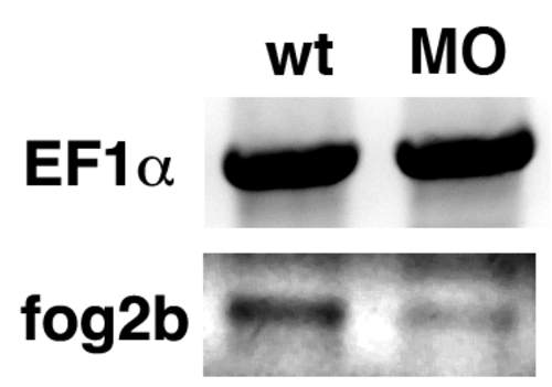
Confirmation of fog2b downregulation in etsrp morphants by RT-PCR analysis. Total RNA from control and morphant embryos at the 16-somite stage was purified and used for cDNA synthesis. EF1? expression was used as a control reference. Primer sequences are listed in Table S1. EXPRESSION / LABELING:
|