
- Title
-
Combination of reverse and chemical genetic screens reveals angiogenesis inhibitors and targets
- Authors
- Kalén, M., Wallgard, E., Asker, N., Nasevicius, A., Athley, E., Billgren, E., Larson, J.D., Wadman, S.A., Norseng, E., Clark, K.J., He, L., Karlsson-Lindahl, L., Häger, A.K., Weber, H., Augustin, H., Samuelsson, T., Kemmet, C.K., Utesch, C.M., Essner, J.J., Hackett, P.B., and Hellström, M.
- Source
- Full text @ Chem. Biol.

Knockdown of ppp1ca, ppp1cc, and ppp4c Results in Defects in Endothelial Path Finding and Tubulogenesis (A–L) Images are all lateral views of the trunk vasculature at 48–50 hpf. (A–C) Control embryo injected with a mixed-base morpholino. (D–L) Embryos injected with morpholinos against (D–F) ppp1ca, (G–I) ppp1cc, and (J–L) ppp4c. Endothelial cells (shown with green fluorescence in [A], [D], [G], and [J]) were identified by using the Tg(fli1:egfp)y1 line. Dorsal aortas are marked with arrowheads, examples of perfused ISVs are marked with arrows, and examples of nonperfused ISVs are labeled with asterisks. (D and J) The ppp1ca and ppp4c knockdowns resulted in enlarged ISVs, whereas (G) knockdown of ppp1cc resulted in excessive branching of the ISVs. (B) At 48–50 hpf, circulation as observed by microangiography was observed in control injected embryos (mixed-base MPO) in the dorsal aorta, cardinal vein, and ISVs. (E and K) Rhodamine-dextran dye (red) often entered the ventral aspect of the ISVs in the ppp1ca and ppp4c knockdowns (a more severely affected embryo is shown for ppp4c), but a circulatory loop was not established. (H) The ppp1cc knockdown embryos showed either an absence of circulation or thin vessels with reduced circulation. (C, F, I, and L) Merged images of the embryos shown in the previous two panels. PHENOTYPE:
|

Treatment of Embryos with Endothall Results in Defects in Tubulogenesis (A, D, and G) Endothelial cells in the trunk were analyzed for defects in migration in the Tg(fli1:egfp)y1 line (green fluorescence) at 48–50 hpf. (B, E, and H) Analysis of circulation defects by using microangiography (rhodamine-dextran imaging, red) at 48–50 hpf. (A–I) are lateral views of the trunk. (A–C) Treatment with either carrier (DMSO control) or (D–I) Endothall did not alter endothelial migration (see Tg(fli1:egfp)y1 in [A], [D], and [G]). (B, E, and H) Microangiography revealed defects is circulation consistent with defects in tubulogenesis. Two classes of affected embryos are shown. In weakly affected embryos (low effect), some of the ISVs would fail to circulate rhodamine-dextran. In more severely affected embryos (high effect), most of the ISVs failed to transfer dye. (C, F, and I) Merged images of the previous two panels. Dorsal aortas are marked with arrowheads, examples of perfused ISVs are marked with arrows, and examples of non-perfused ISVs are labeled with asterisks. (J) Dose response to bathing embryos in Endothall. Embryos were scored based on circulation in the axial vessels and the ISVs as in Table 2. Vascular collapse was observed with the highest concentration of Endothall. PHENOTYPE:
|
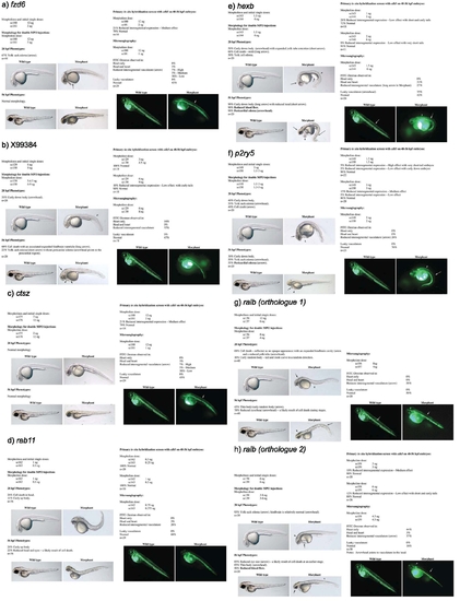
Summary of Zebrafish Knock-Down Phenotypes The results from the MPO knock-down of (a) fzd6, (b) X99384, (c) ctsz, (d) rab11, (e) hexb, (f) p2ry5, (g) ralb, orthologue 1, (h) ralb, orthologue 2, (i) rab5c, (j) ppap2a, (k) ppp4c, (l) ppp1ca, (m) gnb2l1, (n) sat1, (o) ppp1cc, (p) ppih and (q) alox5ap, are listed with regards to: The morphology at 28 and 56hpf of control and MPO knock-down zebrafish embryos. |
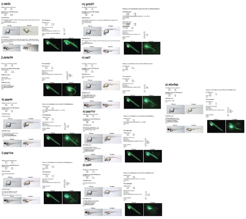
Summary of Zebrafish Knock-Down Phenotypes The results from the MPO knock-down of (a) fzd6, (b) X99384, (c) ctsz, (d) rab11, (e) hexb, (f) p2ry5, (g) ralb, orthologue 1, (h) ralb, orthologue 2, (i) rab5c, (j) ppap2a, (k) ppp4c, (l) ppp1ca, (m) gnb2l1, (n) sat1, (o) ppp1cc, (p) ppih and (q) alox5ap, are listed with regards to: The morphology at 28 and 56hpf of control and MPO knock-down zebrafish embryos. |