
- Title
-
Potential roles of arnt2 in zebrafish larval development
- Authors
- Hill, A.J., Heiden, T.C., Heideman, W., and Peterson, R.E.
- Source
- Full text @ Zebrafish

Arnt2-/- larvae have normal external morphology at 120 hpf. A representative WT larva and arnt2-/- larva are shown. They have similar external morphology, except that swim bladder is not inflated in the mutant. PHENOTYPE:
|
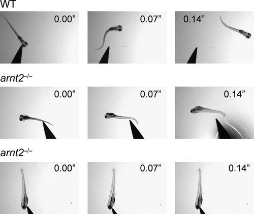
The escape response to a touch stimulus is impaired in arnt2-/- larvae at 120 hpf. When touched on the head a representative WT larva instantly reacts by swimming rapidly away from the mounted probe (top). The same brisk escape response is observed in the WT larva when touched on the tail (results not shown). In contrast, a representative arnt2-/- larva at 120 hpf shows either a very weak response when touched on the tail (middle) or no response when touched on the head (bottom). The video images were recorded at 1 frame/0.07 s after the larva was touched. Results are representative of n=6 WT or arnt2-/- larvae. PHENOTYPE:
|

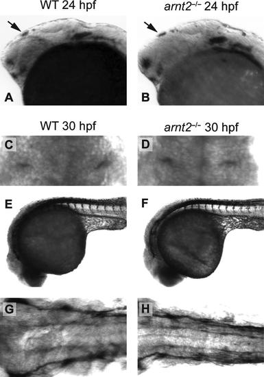
Early brain development appears normal in arnt2-/- embryos at 24–48 hpf. Brain development in the arnt2-/- mutant embryo was assessed by gross examination of the brain at the light microscopic level at 24 hpf. A dorsal view of the developing brain of a WT embryo and an arnt2-/- embryo (A, B) and a lateral view of the brain of a WT embryo and an arnt2-/- embryo (C, D) are shown. At 48 hpf a dorsal view of the brain of a representative WT embryo and arnt2-/- embryo (E, F) is shown. White arrowheads point to ventricles in the brain of the WT and arnt2-/- mutant, demonstrating that brain ventricles are similar in size and shape in the two genotypes (E, F). Overall, gross brain morphology of arnt2-/- larvae at 24–48 hpf was indistinguishable from WT. Patterns of staining at 24 hpf are shown for whole-mount ISH of krox20 in a representative WT embryo and arnt2-/- embryo at 24 hpf (G, H, arrowheads) and of pax6a (I, J, arrowheads). |
Sensory neurons involved in the touch and startle responses appear normal in arnt2-/- embryos at 24–30 hpf. Lateral views for whole-mount ISH of islet1 in a representative WT larva and arnt2-/- larva is shown at 24 hpf (A, B). The pattern of islet1 staining in the brain (black arrow) is similar for the two genotypes, suggesting that regions where primary neurons form are developing normally. 3A10 IHC staining of Mauthner neurons is similar in a representative WT embryo and arnt2-/- mutant embryo at 30 hpf (C, D). ZNP1 IHC to identify primary motor neurons and trigeminal ganglia is shown in a lateral view of a WT embryo and an arnt2-/- embryo (E, F) and in a dorsal view (G, H) at 30 hpf. ZNP1 IHC is similar in the two genotypes. Results in all panels are representative of 5–9 embryos/genotype. |

Morphology of the inner ear, neuromasts, and primary motor neurons appears normal in arnt2-/- larvae at 72–148 hpf. Structures of the inner ear, including the semicircular canals and otolith of a WT larva and an arnt2-/- larva at 72 hpf, are similar (A, B). Neuromasts, the lateral line neurons that sense vibration in the water, are stained with DASPEI and appear as white dots on the lateral surface of WT and arnt2-/- larvae (C, D). The DASPEI-stained neuromasts are similar in number and distribution in the two genotypes. ZNP1-stained primary motor neurons are shown in the trunk of a representative WT larva and arnt2-/- larva at 148 hpf (E, F). Skeletal muscle of the trunk is also shown in these lateral views (E, F). Both ZNP1-stained primary motor neurons and trunk skeletal muscle of WT and arnt2-/- larvae appear to be similar. EXPRESSION / LABELING:
PHENOTYPE:
|
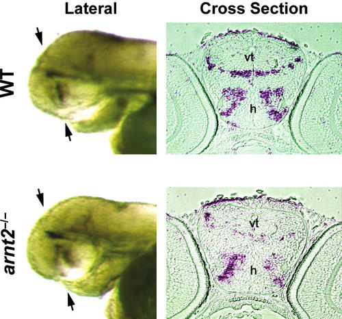
mRNA Expression of the Arnt2 dimerization partner, Sim1, is reduced in the hypothalamus and ventral thalamus of arnt2-/- larvae at 72 hpf. ISH of sim1 in a cross section of the brain taken through the ventral thalamus and hypothalamus of a representative WT larva and arnt2-/- larva at 72 hpf shows reduced expression in the arnt2-/- mutant (compare right top and right bottom). In the left panel, black arrows indicate the plane at which the cross section (shown in the right panel) was taken. Eyes of the representative WT larva and arnt2-/- larva were removed after euthanasia (left top and left bottom) so that the pattern of sim1 staining in the whole-mount brain could be seen in lateral view. vt, ventral thalamus; h, hypothalamus. EXPRESSION / LABELING:
|

Brain ventricle area is increased in arnt2-/- larvae at 120 hpf. Cross section of the brain of a representative WT larva shows a small ventricle to which a black arrow is pointing (top panel). On the other hand, a cross section taken through the same region of the brain of a arnt2-/- mutant reveals a large brain ventricle (black arrow, bottom panel). The two cross sections were taken at similar locations along the anterior–posterior axis of the brain using the tectum, tc, and presumptive pituitary, p (white arrow), for orientation of the sections. h, Hypothalamus; tg, tegmentum; pc, parachordal cartilage. PHENOTYPE:
|

Heart ventricle enlargement, bradycardia, and cardiac arrhythmia in arnt2-/- larvae at 120 and 144–148 hpf. In a representative longitudinal section of the heart (A) of a WT larva (left) and an arnt2-/- larva (right) at 120 hpf, the ventricle of the arnt2-/- mutant appears slightly larger than WT. Transverse sections of the ventricle at 120 hpf were taken along its anterior–posterior axis: anterior (B), middle (C), and posterior (D) for WT (left) and arnt2-/- mutant (right), respectively. A, Atrium; BA, bulbous arteriosus; SV, sinus venosus; V, ventricle. Line graphs (E) show ventricle cross-sectional area as a function of section number (top line graph) and ventricle wall thickness as a function of section number (bottom line graph). WT (line, open circle) and arnt2-/- (line, solid triangle) results shown in the line graphs are for n=7 larvae=genotype at 120 hpf. Mean ventricle area and ventricle wall thickness based on all cross section measurements of the ventricle also are shown for WT and arnt2-/- mutant’s heart (F, bar graphs, right and middle). Heart rate assessed in WT and arnt2-/- larvae at 144 hpf (G) revealed the presence of bradycardia in arnt2-/- larvae (WT, n=4; arnt2-/-, n=11). Asterisk indicates a significant difference (p<0.05). |
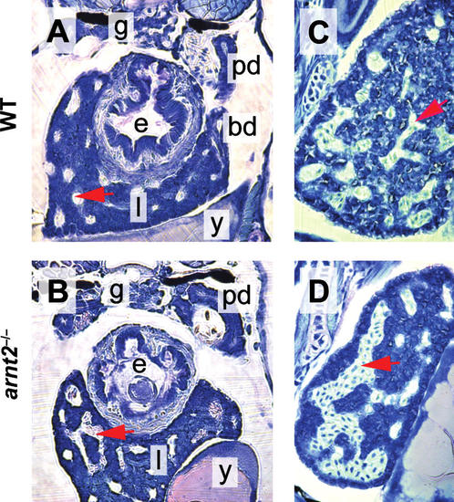
Altered liver sinusoid morphology in arnt2-/- larvae at 120 hpf. Photomicrographs of sections of the liver of a representative WT larva (A, C) and arnt2-/- mutant (B, D) are shown. Compared to WT, the arnt2-/- liver is characterized by enlarged sinusoids engorged with blood that have merged to form large labyrinth-like vascular channels. A red arrowhead points to a blood-filled sinusoid in each of the sections. e, Esophagus; l, liver; y, yolk; g, glomerulus; pd, pronephric duct; bd, bile duct. PHENOTYPE:
|