
- Title
-
T-Cell Factor 4 (tcf7l2) Is the Main Effector of Wnt Signaling During Zebrafish Intestine Organogenesis
- Authors
- Faro, A., Boj, S.F., Ambrósio, R., van den Broek, O., Korving, J., and Clevers, H.
- Source
- Full text @ Zebrafish

apcmcr/mcr fish fail to express some differentiation markers in endoderm-derived tissues. Dorsal view of 72 hpf in wild-type (A, C, E, G) and apcmcr/mcr (B, D, F, H) siblings after WISH for intestinal-fatty acid binding protein (i-fabp) (A, B), trypsin (C, D), liver fatty acid binding protein (l-fabp) (E, F), and insulin (G, H). i-fabp and l-fabp expression is absent in apcmcr mutants, whereas trypsin expression is strongly reduced in these embryos: insulin expression is not affected. Arrowhead indicates ectopic expression of insulin marker in apc mutants (H). EXPRESSION / LABELING:
PHENOTYPE:
|

Endoderm progenitor specification is not affected in apcmcr mutant fish. Dorsal view of 56 hpf in wild-type (A, C, E) and apcmcr/mcr (B, D, F) siblings after WISH for foxA2 (A, B), hhex (C, D), and ptf1a (E, F). The expression pattern of these progenitor markers is not compromised in apc mutant fish. EXPRESSION / LABELING:
|
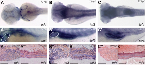
Tcf transcription factors tcf1, tcf3, and tcf4 have a distinctive expression pattern at 72 and 100 hpf. Dorsal view of 72 hpf wild-type fish after WISH for tcf1 (A), tcf3 (B), and tcf4 (C). Detail of lateral view (dorsal to the top) of 72 hpf wild-type fish after WISH for tcf1 (A′), tcf3 (B′), and tcf4 (C′). Histological analysis of sagittal sections of 100 hpf zebrafish larvae after whole-mount in situ for tcf1 (A″, A″′), tcf3 (B″, B″′), and tcf4 (C″, C″′), depicting expression of mRNA in the liver bud and intestinal epithelium, respectively. There is coexpression of tcf1, tcf3, and tcf4 in the intestinal epithelium at these developmental time points. White dashed line delimits domain of expression of tcf1 and tcf3 in liver bud (A′, B′), as confirmed by histological section (A″, B″). Scale bar corresponds to 50 μM. EXPRESSION / LABELING:
|

Tcf4 depletion in apcmcr/mcr background rescues the expression of i-fabp, but not trypsin or l-fabp. Dorsal view of 72 hpf apcmcr/mcr (A, C, E) and apcmcr/mcr;tcf4exI/exI (B, D, F) siblings after WISH for i-fabp (A, B), trypsin (C, D), and l-fabp (E, F). Histological analysis of transversal sections from the foregut of 80 hpf apcmcr/mcr (G) and apcmcr/mcr;tcf4exI/exI (H) larvae stained with hematoxylin and eosin. Apc mutant embryos fail to develop properly polarized enterocytes, whereas in the double-mutant embryos this defect is rescued. EXPRESSION / LABELING:
PHENOTYPE:
|

Compound embryos apcmcr/mcr; tcf4exI/exI display the apc mutant cardiac phenotype. Dorsal view of 48 hpf wild-type (A), apcmcr/mcr (B), and apcmcr/mcr;tcf4exI/exI (C) siblings after WISH for bmp4. Survival of apc tcf4 genotypes at 96 hpf (D). The depletion of Tcf4 in apcmcr/mcr genetic background (gray bars) does not improve the survival of the apc mutant embryos. Values are mean ± standard error of mean; n>25 embryos per category; experiment was triplicated. |
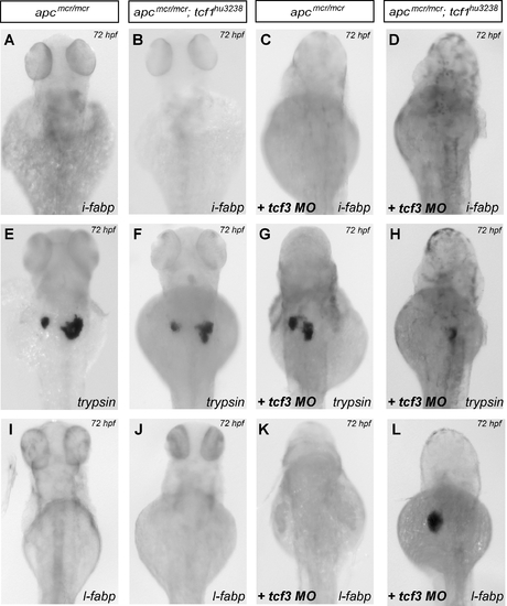
Depletion of either Tcf1 or Tcf3 in apcmcr/mcr genetic background does not rescue expression of differentiation markers in endoderm-derived tissues, but combined depletion recovers expression of l-fabp. Dorsal view of 72 hpf of apcmcr/mcr (A, E, I), apcmcr/mcr;tcf1hu3238/hu3238 (B, F, J), apcmcr/mcr;tcf3MO (C, G, K), and apcmcr/mcr;tcf1hu3238/hu3238;tcf3MO (D, H, L) after WISH for i-fabp (A–D), trypsin (E–H), and l-fabp (I–L). EXPRESSION / LABELING:
PHENOTYPE:
|
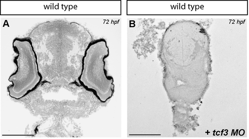
tcf3 MO Injection recapitulate headless phenotype. Histological analysis of transversal sections from the head of 72 hpf wild-type (A) and morphant (B) larvae stained with hematoxylin and eosin. Morphant embryos present severe head formation defects. Scale bar corresponds to 100 μM. PHENOTYPE:
|

apc MO Injection in hdl-/- mutants abrogates the expression of i-fabp but not trypsin. Dorsal view of 56 hpf hdl-/- (A) and hdl-/-;apcMO (B) siblings after WISH for l-fabp and trypsin (A, B). White star denotes trypsin staining in both noninjected and morphant embryos. EXPRESSION / LABELING:
|

Unillustrated author statements PHENOTYPE:
|