
- Title
-
Hardwiring of fine synaptic layers in the zebrafish visual pathway
- Authors
- Nevin, L.M., Taylor, M.R., and Baier, H.
- Source
- Full text @ Neural Dev.
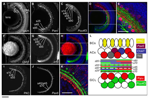
IPL organization of the larval zebrafish retina. Confocal images of horizontal sections of 5 dpf retina stained by immunohistochemistry or with DAPI (nuclear dye). IPL sublaminae are labeled (s10, s25, and so on). (A) DAPI stain shows the basic organization of the retina into GCL, IPL, INL, and ONL. (B-E) Neurites from Parv+ ACs (red) and Pou4f3:mGFP+ GCs (green) are closely apposed but reside in distinct sublaminae. (F-H) Neurites from ChAT+ ACs (red) overlap with zrf3 label (green) in the same sublaminae. (I-K) PKC+ BC axon terminals (red) and Pax6:mGFP+ AC neurites (green) each form three sublaminae that are closely nested but not co-localized. (L) Schematic of IPL organization. Cell types and their neurites are labeled according to the color code on the right. Space is shown between sublaminae for clarity. TH, tyrosine hydroxylase. Bottom left scale bar, for whole retina images, is 50 μm; bottom left scale bar in E, K is 25 μm. |

Neuropil organization of the larval zebrafish optic tectum. Confocal images of horizontal sections of the 5 dpf tectum stained by immunohistochemistry or with DAPI. The neuropil of one lateral half of the tectum is shown. Rostral is up. (A) The Shh:GFP transgene labels all GCs, which innervate the SO, three sublaminae of the SFGS (labeled B, D, and F), the SGC, and the SAC/SPV border. (B) Parv+ tectal neurites form up to five laminae, within the SO, SFGS, SGC, and SAC. The thinnest Parv+ projection is just beneath the skin, superficial to the ShhGFP+ SO projection, likely corresponding to the stratum marginale (SM; most visible in C). (C) Parv+ neurites and GC axons co-localize in the SO and SFGS, but not in the deeper tectal layers. (D) Pou4f3:mGFP+ GC axons label the SO and two sublaminae (labeled D and F) of the SFGS. (E) PKC+ tectal neurites are most dense in three bands in SO, SFGS, and SGC. (F) Schematic showing organization of the tectal neuropil. Scale bar 50 μm. |
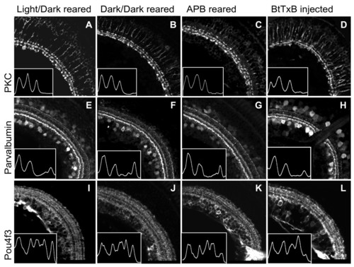
Dark-reared, APB-treated, and BtTxB-injected larvae show proper IPL sublamination.(A-L) Sections showing the IPL of 5 dpf larvae raised in a normal light:dark cycle (A, E, I), constant darkness (B, F, J), in the presence of 1 mM APB (C, G, K), and treated with BtTxB (D, H, L). The images in D, H, L are from the larva recorded in Figure 4D. Insets: traces of the fluorescent signal intensity across the width of the IPL (region shown). Peaks correspond to bands in the IPL. (A-D) PKC+ BC axon terminals are confined to three inner sublaminae in all larvae. (E-H) Parv+ neurites are in three bands in all larvae. The interruption of the IPL in H is the optic nerve. (I-L) Pou4f3:mGFP+ dendrites stratify in five bands in all larvae. Scale bar 50 μm. |

Dark-reared, APB-treated, and BtTxB-injected larvae show proper GC axon targeting to tectal laminae. Horizontal sections of 5 dpf larval tecta showing Pou4f3:mGFP+ GC axons innervating the optic tectum, imaged by confocal microscopy. (A, C, E, G) Pou4f3:mGFP+ axons innervate the SO and two sublaminae of the SFGS. Insets: densitometric traces across the tectal neuropil, from superficial to deeper layers. (B, D, F, H) Same images of Pou4f3+ axons (green), with DAPI labeling (blue) to show the cell body and neuropil regions of the tectum. Scale bar 50 μm. |

Tyrosine hydroxylase-positive neurites innervate the edges of the IPL. Sectioned 5 dpf retina immunostained to tyrosine hydroxylase (TH), imaged by wide-field fluorescence microscopy, shows small processes at the inner plexiform layer edges (arrows). The photoreceptor autofluorescence is particularly high in this image; the photoreceptors are not TH immunopositive. Scale bar 50 μm. |