
- Title
-
Nogo-Nogo receptor signalling in PNS axon outgrowth and pathfinding
- Authors
- Brösamle, C., and Halpern, M.E.
- Source
- Full text @ Mol. Cell Neurosci.
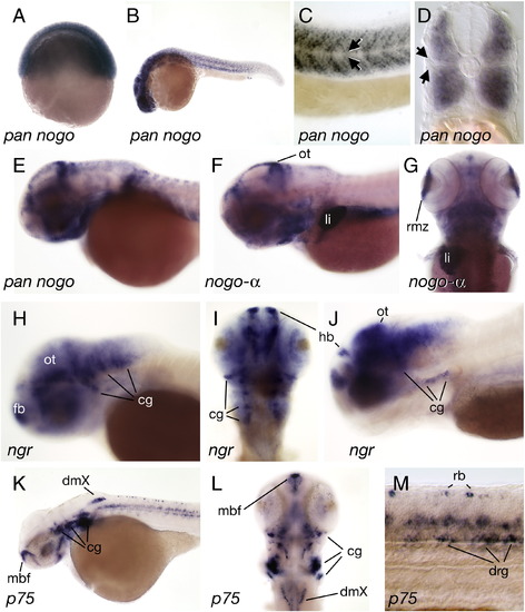
Expression of nogo, nogo receptor, and p75 in the developing zebrafish. (A) nogo transcripts were detected as early as 6 hpf. (nogo-α was not detected at this stage, not shown). (B) At 24 hpf, nogo was expressed in many neural and non-neural tissues but (C, D) expression was absent from a discrete domain (arrows) between the dorsal and ventral trunk muscles. (E, F, G) At 2 and 3 dpf, nogo transcripts were abundant in the posterior rim of the optic tectum (ot) and the marginal zone of the retina (rmz). Strong nogo-α expression was also detected in the liver starting at 3 dpf. (H) at 36 hpf and (I, J) 4 dpf ngr was expressed in many neural populations including cells in the forebrain (fb), habenula (hb), optic tectum, and cranial ganglia (gc). (K–M) Gene expression for the low-affinity neurotrophin receptor p75 was detected in the medial basal forebrain (mbf), cranial ganglia, dorsal motor nuclei of the vagus (dmX) and, at lower levels, in the retina at 2 dpf. (M) In the spinal cord, p75 was expressed in Rohon-Beard neurons (rb) and dorsal root ganglion neurons (drg). (A–C, E, F, H, J, K, M) side views, (D) cross section, (G, I, L) dorsal views. |
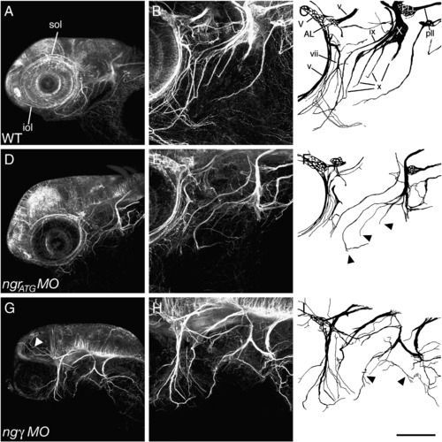
Loss of Nogo-γ or Ngr function disrupts cranial nerve projections. (A–C) Anti-acetylated tubulin immunostaining at 3 dpf reveals highly stereotyped innervation of the larval head, allowing the identification of many cranial nerves (Higashijima et al., 2000). Injection of 5 ng of (D–F) ngrATG MO or (G–I) nogo-γ MO results in the disruption of the cranial innervation pattern. Many nerve branches were significantly thinner, shorter, or even absent. In addition, nerve trajectories were disturbed and axons projected into aberrant target areas (arrowheads). (A, B, D, E, G, H) Maximum intensity projections of side view confocal z-series of whole-mount zebrafish larvae. (B, D, F) are higher magnifications of the area caudal to the eye shown in (A, C, E), and depicted schematically in (C, F, I). iol = infraorbital lateral line nerve, sol = supraorbital lateral line nerve, V = trigeminal ganglion, v = trigeminal nerve, AL = anterior lateral line ganglion, vii = facial nerve, ix = hypoglossal nerve, X = vagal ganglion, x = branches of the vagal nerve, PLL = posterior lateral line ganglion, pll = posterior lateral line nerve. Scale bar A, C, E = 200 μm; B, D, F = 100 μm. PHENOTYPE:
|
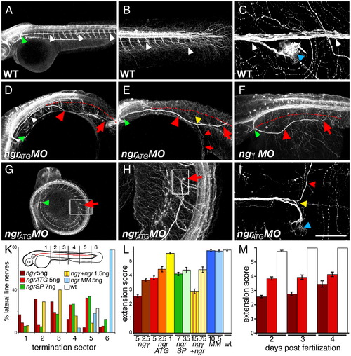
Posterior lateral line (pll) nerve defects result from Nogo-γ and Ngr depletion. (A) Bilaterally, the pll nerve emerges from the pll ganglion (indicated by green arrowhead) and projects caudally along the trunk to reach the ventral base of the tail by the end of 2 dpf (white arrowheads, B). (C) As it traverses the length of the embryo, pll axons innervate sensory organs of the lateral line (blue arrowhead). In (D, E) ngrATG, (F) nogo-γ, and ngrSP (not shown) morphants, the trajectory was frequently abnormal (indicated by red arrowheads), with the pll nerve veering off the correct path (red dotted line), invading inappropriate areas and terminating prematurely (the end of the nerve is indicated by red arrows). (E) In some instances, the pll nerve was found to branch (yellow arrowhead). (G–I) Additionally, many pll nerves terminated prematurely (G–I, H and I higher magnifications of boxed areas in G and H, respectively). NgrATG morphants were frequently curled ventrally (G) and anterior is indicated by (a). (K) The extent of pll outgrowth was scored according to the embryonic sector in which the pll nerve terminated at 48 hpf. In control WT or mismatch morpholino injected larvae, > 95% pll nerves reached sector 5 or 6 (white and blue bars). Following MO injections for ngrATG (red), (green), nogo-γ (brown), or a combination of ngrATG and nogo-γ (striped bars) only between 11% (nogo-γ) and 44% (ngrSP) of nerves terminated in sectors 5 and 6. (L) Average extension scores indicated that pll truncation was dose-dependent and that co-injection of ngr and nogo-γ morpholinos potentiated the effect. (M) Incomplete extension of the pll nerve persisted at 3 and 4 dpf. (A–I) Maximum intensity projections of side view confocal z-series of 2 dpf anti-acetylated tubulin immunostained whole-mount zebrafish larvae. Scale bar A and G 400 μm; B, and D–F 200 μm; H 50 μm; C and I 40 μm. |

Unillustrated author statements PHENOTYPE:
|
Reprinted from Molecular and cellular neurosciences, 40(4), Brösamle, C., and Halpern, M.E., Nogo-Nogo receptor signalling in PNS axon outgrowth and pathfinding, 401-409, Copyright (2009) with permission from Elsevier. Full text @ Mol. Cell Neurosci.