
- Title
-
Isolation and expression analysis of Alzheimer's disease-related gene xb51 in zebrafish
- Authors
- Kim, H.T., Kim, E.H., Yoo, K.W., Lee, M.S., Choi, J.H., Park, H.C., Yeo, S.Y., Lee, D.S., and Kim, C.H.
- Source
- Full text @ Dev. Dyn.
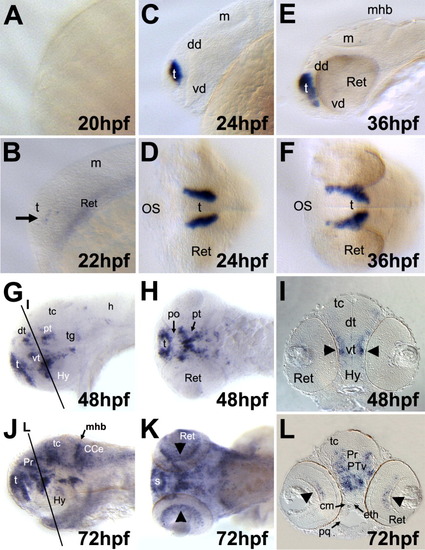
Spatiotemporal expression pattern of the xb51 gene in zebrafish embryos. Lateral views in A-C, E, G and J. Frontal views in D and F. Dorsal views in H and K. Anterior to the left (A-C, E, G, H, J, and K). A: At 20 hpf, xb51 transcripts were not detected in any embryonic region. B-F: xb51 expression was detected in forebrain from 22 hpf (arrow in B). At 24 hpf, the bilateral domains of xb51 expression were detected (C, D); its expression increased in the forebrain by 36 hpf (E, F). G-I: At 48 hpf, xb51 expression is extended to the diencephalon (G, H). In cross-section of the 48-hpf embryo, the diencephalic expression domains were present laterally close by pial surface (I, arrowheads). J-L: At later stages, xb51 expression domains extend to the midbrain, hindbrain, and retina (arrowheads). CCe, cerebellum; cm, craniofacial mesenchyme; dd, dorsal diencephalon; eth, ethmoid plate; dt, dorsal thalamus; h, hindbrain; Hy, hypothalamus; m, midbrain; mhb, midbrain-hindbrain boundary; OS, optic stalks; po, preoptic area; pq, palatoquadrate; Pr, pretectum; pt, posterior tuberculum; PTv, ventral part of the posterior tuberculum; ret, retina; s, subpallium; t, telencephalon; tc, tectum; tg, tegmentum; vd, ventral diencephalon; vt, ventral thalamus. EXPRESSION / LABELING:
|
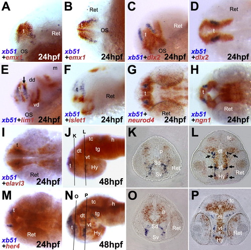
Double in situ hybridization of xb51 and with several molecular markers. Front-lateral (A, C, E, F), dorsal (B, D, G, H, I, M), and lateral views (J, N). Anterior to the left. A-D: At 24 hpf, the expression domain of xb51 is overlapped with that of emx1, a dorsal telencephalon marker (A, B), but not with dlx2, a ventral telencephalon and diencephalon marker (C, D). E-H: The expression domain of xb51 is partly overlapped with that of lim1 in the dorsal telencephalon at 24 hpf (arrow in E), while it is not overlapped with that of islet1 (F), neurod4/zath3 (G), and ngn1 (H). I-L: At 24 hpf, the xb51 expression domain is included in that of elavl3/huC, a pan-neuronal marker (I). At 48 hpf, in cross-section (J-L), the expression domain of xb51 is overlapped with elavl3/huC in the lateral region of the brain (arrows). M-P: The xb51 expression domain is not overlapped with that of her4, a marker for proliferating undifferentiated neural cells. In cross-sections of embryos at 48 hpf (O, P), expression domains for xb51 and her4 show a complementary pattern for each other. Approximate planes of the sections are indicated by lines in J and N. p, pallium; Sd, dorsal subpallium; Sv, ventral subpallium. |

Expression of xb51 in neuronal cells in the forebrain. Lateral (A-C, G, H, K, L), frontal (D-F), and dorsal views (I, J). Anterior to the left (A-C, G, H, K, L). A, D: Embryo hybridized with an xb51 riboprobe at 30 hpf (A) and 36hpf (D). B, E: Embryo stained with an anti-acetylated α-tubulin antibody, showing primary axon tracts. C, F: Double staining of embryos with xb51 and the anti-acetylated α-tubulin antibody. The xb51-expressing domain is very close to the region from where the anterior commissural axons (ac) originate (arrows). G-L: Expression of xb51 in a neurogenic mutant, mind bomb. The bilateral expression domains of xb51 in wild-type embryos (I) are medially fused in mind bomb mutant embryos at 24 hpf (J). At 36 hpf, xb51-expressing cells are ectopically detected in the midbrain of mind bomb mutant embryos (arrow). ac, anterior commissure; mlf, medial longitudinal fasciculus; NTAC, nucleus of the tract of the AC; po, posterior commissure; poc, postoptic commissure; sot, supraoptic tract; tpoc, tract of the postoptic commissure. |