
- Title
-
Nodal signaling promotes the speed and directional movement of cardiomyocytes in zebrafish
- Authors
- de Campos-Baptista, M.I., Holtzman, N.G., Yelon, D., and Schier, A.F.
- Source
- Full text @ Dev. Dyn.
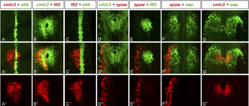
Expression of Nodal pathway genes in the lateral plate mesoderm. Confocal images of double fluorescent in situ RNA hybridization of spaw, lft2, and oep with respect to the midline marker shh and the heart marker cmlc2 at the 21-somite stage. Single channel expression (A-G and A″-G″) and overlay (A′-G′). All images are dorsal views of the embryo, anterior to the top and left to the left. Note broad expression of oep in left and right LPM (F and G) and more restricted expression of spaw in left non-cardiac LPM (F′ and F″) and lft2 in left cardiac LPM (B′ and B″). spaw is expressed adjacent to lft2 (E′). EXPRESSION / LABELING:
|
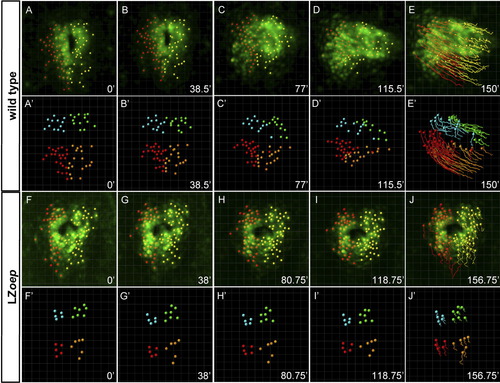
Tracking of single cells in wild type and LZoep mutants. Confocal 3D-images showing individual cmlc2-GFP cells that were manually tracked starting at the onset of cardiac cone formation. First (A-E) and third (F-J) rows: green cmlc2-GFP expression; red: cells within the left side of cardiac cone; yellow: cells within the right side of cardiac cone. For detailed views from different angles, see Supplemental Movies 1 (wild type) and 5 (LZoep). Second (A′-E′) and fourth (F′-J′) rows: tracking of quadrants. Quadrants were defined by grouping left-anterior cells (blue), left-posterior cells (red), right anterior cells (green), and right-posterior cells (orange). For detailed views from different angles, see Supplemental Movies 3 (wild type) and 7 (LZoep). Pictures were cropped to keep the heart in the center and do not maintain the position of the coordinate systems. For movements with respect to coordinate systems, see Supplemental Movies 3 (wild type) and 7 (LZoep). EXPRESSION / LABELING:
|
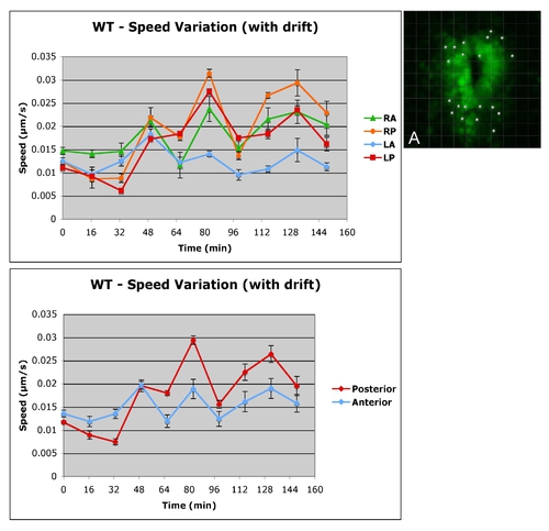
Comparison of tracking strategies. Quantitative analysis of anterior and posterior cell movements in wild type similar to Smith et al. (2008), i.e., without correction for drift of the embryo during imaging or for displacement of heart within the embryo. Note that speed differences between posterior and anterior cells are maintained as in the analysis that uses the base of the lumen as a reference point (Figs. 3-5), but speed values are higher, in agreement with Smith et al. (2008). |