
- Title
-
Eya4 regulation of Na+/K+-ATPase is required for sensory system development in zebrafish
- Authors
- Wang, L., Sewell, W.F., Kim, S.D., Shin, J.T., Macrae, C.A., Zon, L.I., Seidman, J.G., and Seidman, C.E.
- Source
- Full text @ Development
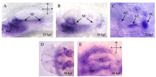
eya4 expression during otic vesicle development. Wild-type embryos were hybridized to DIG-labeled eya4 antisense RNA probes. Lateral (A-C) and dorsal (E) views of single otic vesicles; (D) dorsal view of two otic vesicles. eya4 signal (arrows) at 24 hpf (A) and 36 hpf (B) demarcated the developing sensory epithelia (SE) and neuroblast (NB). At 72 hpf (C), expression was maintained in the anterior, lateral and posterior cristae (AC, LC and PC, respectively) of the sensory epithelia. At 48 hpf (D,E), eya4 signal was more diffuse within the otic vesicle and was prominent in the anterior macula (AM) and pharyngeal arch (PA; E). Background signal with eya4 sense RNA probe was minimal (data not shown). D, dorsal; V, ventral; A, anterior; P, posterior; M, medial; L, lateral. EXPRESSION / LABELING:
|
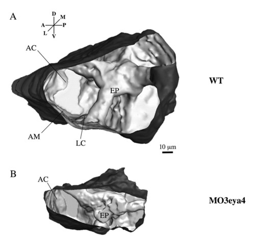
Three-dimensional reconstruction of the zebrafish otic vesicle (72 hpf) generated from confocal images. (A) The otic vesicle of the wild-type zebrafish has well-formed epithelial pillars (EP) that shape the semicircular canals. The anterior cristae (AC), lateral cristae (LC) and anterior maculae (AM) are indicated. (B) The otic vesicle of an eya4 morphant fish is smaller and misshapened, with fused epithelial pillars and malformed semicircular canals. PHENOTYPE:
|
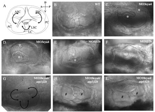
Otic vesicle morphology in wild-type and morphant fish (72 hpf). (A) Schematic of the normal otic vesicle structures visualized by differential interference contrast (DIC) imaging. Anterior, lateral and posterior cristae (AC, LC and PC, respectively) have differentiated into anterior, lateral and posterior semicircular canals (ASC, LSC and PSC, respectively) that are portioned by epithelial pillars (EP) inside the otic vesicle. (B) DIC image of the otic vesicle in wild-type fish reveals normal structures. (C,D) DIC images of otic vesicles from eya4 morphant fish show aborted protrusions of presumptive epithelial pillars (asterisks) into the vesicle and malformed canals. (E,F) DIC images of atp1b2b morphant fish otic vesicles show incomplete fusion and diminutive epithelial pillars (asterisks), which resemble those of the eya4 morphants. (G-I) atp1b2b mRNA-rescued eya4 morphant fish otic vesicles were still smaller than wild type, but were larger than eya4 morphant vesicles. Canal formation was partially (# in H,I) or nearly completely restored (G) as a result of the more mature epithelial pillars. PHENOTYPE:
|
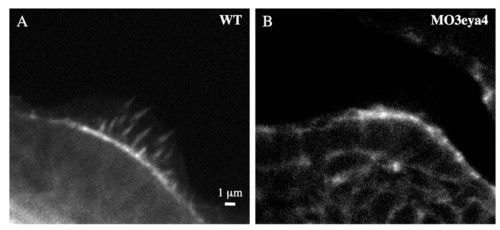
Compromised hair cell development eya4 morphant fish. (A) At 72 hpf the ampulla of the posterior cristae of wild-type fish (n=10) have delicate hair cell stereocilia (stained with FITC-labeled phalloidin) that appear as spikes. (B) Stereocilia in hair cells of eya4 morphant fish (n=12) are dysmorphic and reduced in number. PHENOTYPE:
|
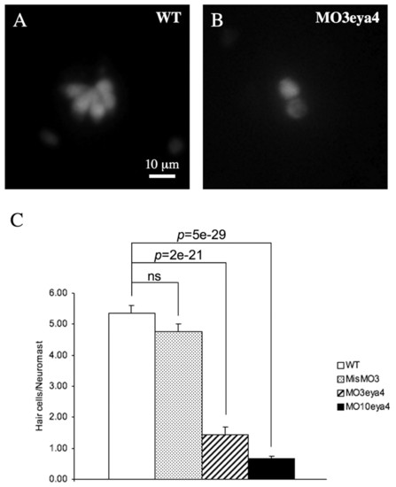
Reduced numbers of neuromast hair cells in eya4 morphant fish. Wild-type (A) and eya4 morphant (B) fish were stained with DASPEI, and neuromast hair cells were imaged and counted (C). PHENOTYPE:
|
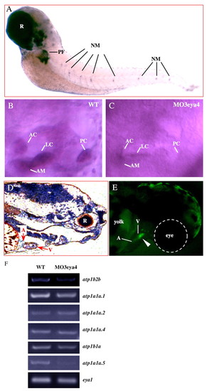
atp1b2b expression in zebrafish embryos (72 hpf). (A) Whole-mount in situ hybridization with DIG-labeled atp1b2b antisense probe revealed expression in the otic vesicle, neuromast (NM), retina (R) and pectoral fin (PF). (B) Higher resolution image of atp1b2b expression in a lateral view of the otic vesicle with robust signals in the anterior macula (AM), and the anterior, lateral and posterior cristae (AC, LC and PC, respectively) in wild type. (C) In the eya4 morphant otic vesicle, atp1b2b expression was notably reduced in the AC, LC, PC and AM. (D) In situ hybridization of sections using a radiolabeled atp1b2b antisense probe show atp1b2b expression in the heart. Expression in the ventricle (V) is greater than in the atrium (A). DIG and radiolabeled atp1b2b sense probes produced low background signals (data not shown). (E) Fluorescent signal (arrowhead) detected at 72 hpf in zebrafish injected with plasmid pβ2bprom confirmed atp1b2b promoter activity and EGFP expression. (F) Semi-quantitative RT-PCR (see Materials and methods) showed reduced atp1b2b expression in eya4 morphant fish compared with wild type. MO3eya4 did not alter eya1 expression (Schonberger et al., 2005). Semi-quantitative RT-PCR on other Na+/K+-ATPase subunits that were expressed in the otic vesicle, including atp1a1a.1, atp1a1a.2, atp1a1a.4, atp1b1a and atp1a1a.5, are also shown. EXPRESSION / LABELING:
|
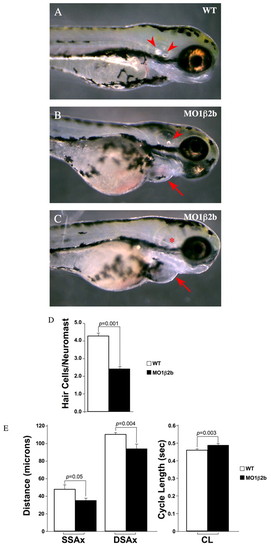
Phenotypes of atp1b2b morphant fish (72 hpf). (A) Otic vesicles from wild-type embryos contained two otoliths (arrowheads). (B,C) atp1b2b morphant fish lacked one (B) or both (asterisk, C) otoliths and had ventral protuberances due to pericardial effusion (arrows), consistent with cardiac failure. (D) atp1b2b morphant neuromasts had significantly fewer hair cells than wild-type embryos. (E) Cardiac ventricular chamber sizes were smaller and contractile cycle length was longer (corresponding to slower heart rate) in atp1b2b morphant fish compared with wild-type fish. SSAx, systolic short axis; DSAx, diastolic short axis; CL, cycle length. PHENOTYPE:
|
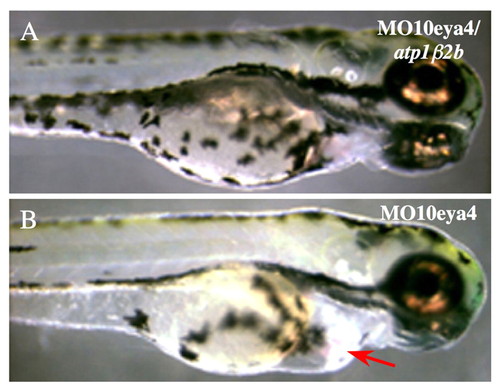
atp1b2b mRNA rescued eya4 morphant phenotypes. (A,B) The hearts of atp1b2b mRNA-rescued eya4 morphant fish (A) lacked pericardial effusion, visible in eya4 morphant fish (arrow, B). PHENOTYPE:
|
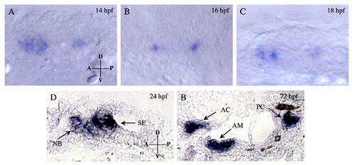
eya4 expression during otic placode and otic vesicle development. (A-C) At 14, 16 and 18 hpf, wild-type embryos were hybridized to DIG-labeled eya4 antisense RNA probes. Lateral views of the otic placode region are shown. (D,E) Sections from wild-type embryos were hybridized with a DIG-labeled eya4 antisense RNA probe. eya4 signal (arrows) at 24 hpf (D) demarcated the developing sensory epithelia (SE) and neuroblast (NB). A section from 72 hpf (E) demonstrated eya4 expression in the anterior maculae (AM), anterior cristae (AC) and posterior cristae (PC) of the sensory epithelia. D, dorsal; V, ventral; A, anterior; P, posterior. EXPRESSION / LABELING:
|
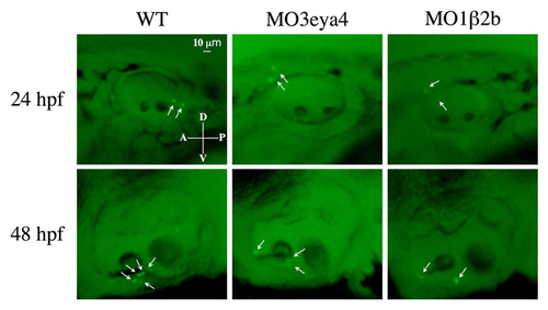
Acridine Orange staining of otic vesicles from 24 and 48 hpf wild-type, eya4 and atp1b2b morphant fish. Positive signals (indicated by white arrows) were observed in a small number of embryos. Those embryos with maximal staining from each group at both time points are presented. In general, only background levels of Acridine Orange staining were observed in wild type, and eya4 and atp1b2b morphants. |

Unillustrated author statements PHENOTYPE:
|