
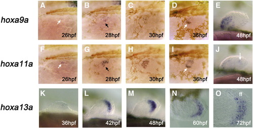
Expression of hoxa9a, hoxa11a, and hoxa13a during zebrafish pectoral fin development. Expression of hoxa9a (A–E) and hoxa11a (F–J) is largely confined to the prospective myogenic cells of the fin bud, whereas expression of hoxa13a (K–O) is seen exclusively within the distal mesenchyme cells that later give rise to the connective tissues of the fin blade. Oblique dorsal (A–D, F–I), lateral (E, J, K–M), or dorsal (N, O) views with anterior to the left in all panels. Only left side is shown for each embryo. Some embryos are also stained for muscle myosin (brown staining in panels A–D, F–I) to show the position of myotomes. Arrows in panels A/F, B/G, and D/I mark the expression within the nascent myogenic cells (A, F), myogenic cells invading the fin bud proper (B, G), and lateral cluster of prospective pectoral fin muscle cells (D, I), respectively. ff: fin fold (later, fin blade). hpf: hours post fertilization. EXPRESSION / LABELING:
|
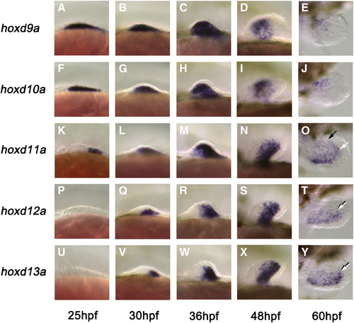
Expression of hoxda cluster genes during pectoral fin development in zebrafish. Expression of hoxda genes occurs in three phases during pectoral fin bud development. Phase I, which occurs at the beginning of fin bud morphogenesis, is characterized by a uniform expression of hoxd9a within the early fin bud mesenchyme (A, F, K, P, U). Phase II, which immediately follows phase I (and for hoxd10a (F), occurs simultaneously with phase I), is characterized by a sequential activation of hoxd10–13a gene expression, each of which occupies successively smaller regions within the fin bud centered at the posterior margin (“early” phase II: B, G, L, Q, V). During later stages (“late” phase II) distal expression domains of these genes extend variably into more anterior regions, thereby causing an appearance of the distal bending of the expression (C, H, M, R, W). Phase III, which is limited to the distal fin bud mesenchyme cells, occurs last and is characterized by the posteriorly limited expression of hoxd11–13a genes within the most distal group of cells (O, T, Y). Note that, unlike Hoxd13 in tetrapod limbs, expression of hoxd13a in zebrafish pectoral fins during this phase does not cover the entire distal fin bud region. During phase III, no expression is seen for hoxd9a and hoxd10a in distal cells (E, J). Lateral (A–D, F–I, K–N, P–S, U–X) or dorsal (E, J, O, T, Y) views with anterior to the left in all panels. Only left pectoral fin buds are shown. Black and white arrows in panels O, T, Y show the anterior limits of expression within the proximal and distal mesenchyme cells, respectively. hpf: hours post fertilization. |
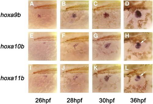
Early expression of hoxab cluster genes in zebrafish pectoral fins. Expression of hoxa9b starts within the mesenchyme cells of anterior fin field (lying lateral to the second myotome) and subsequently spreads toward the posterior, eventually encompassing the entire fin bud mesenchyme by 30 hpf (A–C: phase I expression). For hoxa10b and hoxa11b, the initial expression is confined to the myogenic mesenchyme cells (E–G, I–K). For these genes, expression within the chondrogenic mesenchyme cells begins around 30 hpf in distal posterior cells (phase II expression) roughly overlying the medial group of myogenic cells (H, L: arrows). In dorsal views, this creates a misleading impression of enhanced expression in the medial cluster of prospective pectoral fin muscle cells (compare Figs. 4H, L with Figs. 2D, I). Oblique dorsal views with anterior to the left in all panels. Each embryo is also counter-stained for muscle myosin (brown) to show the position of myotomes. hpf: hours post fertilization. |
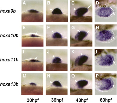
Late expression of hoxab cluster genes during zebrafish pectoral fin development. Similar to hoxda genes, expression of hoxab genes occurs in three distinct phases during pectoral fin bud development. Phase I, which gradually occurs during early stages of fin bud morphogenesis, is characterized by a uniform expression of hoxa9b within the early fin bud mesenchyme (A). Phase II expression begins with the onset of hoxa10/11/13b expression within the distal posterior fin bud mesenchyme just underneath the surface ectoderm (E, I, M), which establishes nested domains of expression for hoxa9–11b genes during later stages (B, F, J, C, G, K). Note that, for hoxa13b (N, O), colinearity is not observed, even though its expression is otherwise similar to the phase II expressions of hoxa10b/a11b during the same period. Phase III, which is limited to the distal fin bud mesenchyme cells, occurs last and is characterized by the expression of hoxa11b and hoxa13b genes within distal cells (L, P: arrows). During phase III, little expression is seen for hoxa9b and hoxa10b genes in the same region (D, H: arrows). Lateral (A–C, E–G, I–K, M–O) or dorsal (D, H, L, P) views with anterior to the left. Only left pectoral fin buds are shown. Arrowheads in panels B, C, F, G, J, K, N, and O show the anterior limits of expression within the chondrogenic mesenchyme cells which at these stages are flanked on both sides by myogenic cells which tend to show weaker expression. Small dots in panels D and H represent the outermost extent of the distal mesenchyme cells. hpf: hours post fertilization. |
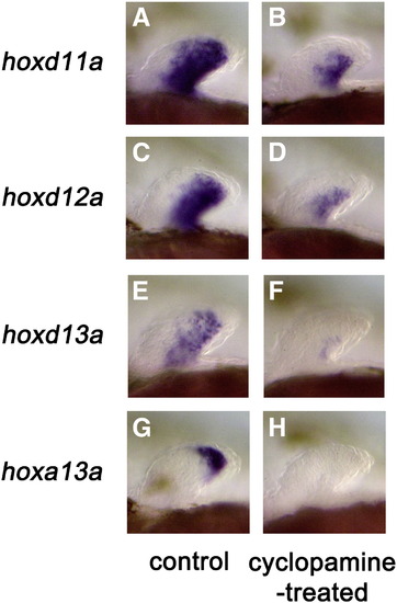
Effect of cyclopamine treatment on hox gene expression in the pectoral fin bud. (A, C, E, G) Control embryos showing normal expression of hox genes. (B, D, F, H) Embryos treated with 50 μM cyclopamine for 12 h from 36 hpf. Note the complete absence of hox gene expression within the distal mesenchyme cells in cyclopamine-treated embryos. Lateral views of the left pectoral fins with anterior to the left in all panels. All embryos are approximately at 48 hpf. hpf: hours post fertilization. EXPRESSION / LABELING:
|
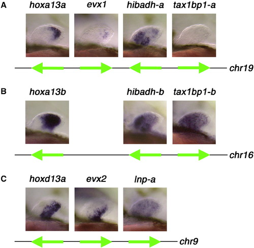
Expression of the genes neighboring the hoxa and hoxd clusters during zebrafish pectoral fin development. (A) Expression of the genes near the 5′ end of the hoxaa cluster. For evx1 and hibadh-a, strong expression is clearly seen within the distal region of the fin bud in a pattern similar to hoxa13a. For tax1bp1-a, little expression is seen except for a small subset of specimens (about 5%; not shown), which show an elevated level of expression in the distal region of the fin in a pattern similar to hoxa13a. (B) Expression of the genes near the 5′ end of the hoxab cluster. Similar to hoxa13b, both hibadh-b and tax1bp1-b are expressed in both proximal and distal regions of the fin bud. (C) Expression of the genes near the 5′ end of the hoxda cluster. For evx2, expression in distal regions is confined to the posterior, which is similar to its 3′ neighbor, hoxd13a. For lnp-a, low level expression is seen throughout the fin bud mesenchyme. Lateral views of left pectoral fin buds with anterior to the left in all panels. All embryos are at 48 hpf. Strong proximal staining of hibadh-a (A) and hibadh-b (B) within the fin bud represents expression within the myogenic cells. The diagram below each set of photographs represents arrangement of genes on the zebrafish genome and the direction of transcription for each gene. Distances between genes are not drawn in scale. For all genes except lnp-a, linkages and precise syntenic relationships are independently confirmed by examining large genomic clones covering the corresponding region. For lnp-a, synteny is presumed based upon its map position in radiation hybrid panels (ZFIN database) and the relative direction of transcription is tentative. chr: chromosome. |
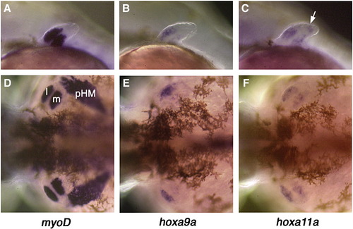
Late expression of hoxa9a/a11a in pectoral fin muscle cells in zebrafish. By 48 hpf, pectoral fin muscle cells can be recognized by the expression of several marker genes such as myoD (A, D), which is not expressed during the migration of fin muscle cells (24–30 hpf: not shown). At this stage, it is clear that the expression of hoxa9a (B, E) and hoxa11a (C, F) is mostly confined to the pectoral fin muscle cells. Note that, for hoxa9a, expression is much weaker in the medial cluster of muscle cells compared to the lateral cluster of muscle cells. For hoxa11a, expression is also seen in the distal mesenchyme cells (arrow in B) in addition to the pectoral fin muscle cells. A–C: lateral view of left pectoral fins. D–F: dorsal-posterior view of pectoral fins and surrounding regions. l: lateral cluster of fin muscle cells. m: medial cluster of fin muscle cells. pHM: posterior hypaxial muscle. EXPRESSION / LABELING:
|

Unillustrated author statements EXPRESSION / LABELING:
|
Reprinted from Developmental Biology, 322(1), Ahn, D., and Ho, R.K., Tri-phasic expression of posterior Hox genes during development of pectoral fins in zebrafish: Implications for the evolution of vertebrate paired appendages, 220-233, Copyright (2008) with permission from Elsevier. Full text @ Dev. Biol.