
- Title
-
Bucky ball functions in Balbiani body assembly and animal-vegetal polarity in the oocyte and follicle cell layer in zebrafish
- Authors
- Marlow, F.L., and Mullins, M.C.
- Source
- Full text @ Dev. Biol.

bucky ball is required to localize vegetal pole and restrict animal pole localized oocyte transcripts. Animal pole localization of pou2 in in situ hybridizations on sections of wild-type stage II (A) and stage IV (B) oocytes. (C) In situ hybridizations on sections show pou2 enriched at multiple cortical sites in buc stage II oocytes, and panel D radially expanded at the cortex in buc stage IV oocytes. (E) Animal pole (AP) view of pou2 localization in a wild-type stage IV oocyte. (F) In buc mutant oocytes pou2 transcripts are not asymmetrically localized. (G) Vg1 localization (arrow) at the animal pole beneath the micropyle in wild-type, and panel H in multiple, ectopic domains in buc mutant oocytes. In situ hybridization on sections reveals brl enrichment at the vegetal cortex (arrowhead) in wild-type early stage II (I) and late stage II (J) oocytes. At stage III of oogenesis, brl transcripts are discretely localized to the vegetal cortex in wild-type (K). In stage II bucky ball mutant oocytes, brl remains throughout the oocyte (L, M) and is also not localized in stage III (N) oocytes. (O) mago nashi transcripts localize to the vegetal pole in wild-type stage III oocytes (arrowhead). (P) Although present in primary oocytes (arrow, also in panel O), mago nashi is not detectable by in situ hybridization in stage III buc oocytes. (A–D, I–N) Hybridizations on slides containing 10 μM ovary sections. (E–H and O–P) Whole mount in situ hybridization images. Stage is shown in the lower, left of each panel. Bars are 50 μm. |
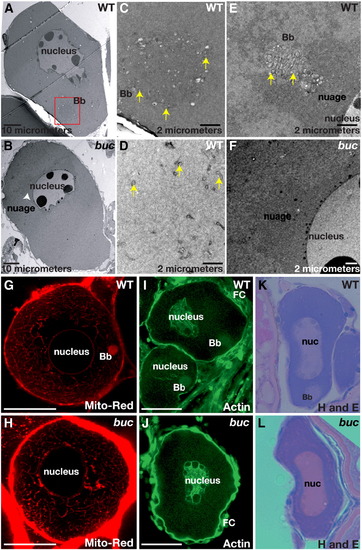
bucky ball is required for Balbiani body assembly. (A, B) 1500x TEM images reveal the Balbiani body (red box) in wild-type (A), but not in buc (B). Mitochondria (yellow arrows) are abundant and clustered within the Balbiani body (C), whereas on the opposite side of the nucleus they are sparse (D, yellow arrows). (E) TEM reveals mitochondria (yellow arrowheads) and associated nuage in the Balbiani body of another wild-type oocyte. Nuage is adjacent to the nucleus and surrounds the wild-type Balbiani body. In buc stage I oocytes, perinuclear nuage is present (F and white arrowhead in panel B). (G) Mitotracker red labels mitochondria in the Balbiani body (Bb) of wild-type stage I oocytes, and (H) reveals abundant mitochondria in buc stage I oocytes, which fail to aggregate into a Bb. (I) Actin localizes to the oocyte nucleus, the somatic cells (FC) and the Bb in wild-type stage I oocytes. (J) In buc mutants, Actin localization is like wild-type except for the lack of a Bb aggregate. Hematoxylin and Eosin stained sections of primary oocytes confirm the presence of a Bb in wild-type (K), but not in buc (L). (C–F) 10,000x TEM; (G–H) 63x confocal images; (I–J) 40x confocal images; (K, L) 40x images. Scale bars are 50 μm for panels G–J. PHENOTYPE:
|

bucky ball ovaries are grossly normal. (A) Wild-type and (B) buc mutant ovaries from adults are of similar size and composition (green circles outline single late stage III or IV oocytes), except that older buc mutant ovaries often have a mass of tissue in the posterior region of the ovary (blue brackets highlight the extent of the posterior mass). Other aspects of oogenesis (oocyte stages I–IV) appeared normal in buc mutants, as revealed by H and E stained adult ovary sections from (C) wild-type and (D) buc. (A, B) Dissecting microscope images. (C, D) 10x images. CG indicates cortical granules located centrally in young oocytes (e.g. box) and at the cortex (arrow) in late stage WT and mutant oocytes. n indicates the oocyte nucleus. Scale bars, 200 μm (A, B); 50 μm (C, D). PHENOTYPE:
|

Asymmetric localization of dazl and Gasz to the Balbiani body fails in bucky ball mutants. (A) In wild-type primary oocytes dazl mRNA localizes to the Balbiani body, (B) then expands toward the vegetal pole. In stages II (C), III (D), and IV (E) wild-type oocytes, dazl transcripts are localized to the vegetal cortex. (K) In wild-type eggs before activation, dazl is localized to the vegetal pole. (F) In bucky ball mutants dazl fails to localize to the Balbiani body in primary oocytes and (G) remains dispersed throughout early stage II oocytes. In stage II (H), early stage III (I), and stage IV bucky ball oocytes (J), dazl mRNA is not localized, although small foci of transcripts are present (yellow asterisks in panel J) at all stages of oogenesis. Arrows indicate the cortex in panels E, J. dazl mRNA remains unlocalized in mutant eggs (L). (M) Hermes protein in wild-type stage I oocytes (pink arrows indicate the Balbiani body). Hermes does not localize in a cytoplasmic aggregate in bucky ball mutant primary oocytes (N). (O) Gasz protein localizes to an aggregate in stage I wild-type oocytes, colocalizing with mitochondria labeled with DiOC6 (P) in the Balbiani body (Q, merged; Gasz (red), DiOC6 (green)). (R) Gasz protein is not asymmetrically localized in bucky ball stage I oocytes. The oocyte stage is shown at the lower right of panels. VE — vitelline envelope. Panels A–J are 5 μM sections following whole mount in situ hybridization. Panels K, L are whole mounts. (M–R) Confocal images (63x objective). Bars = 50 μm. |
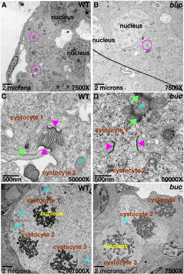
Cytoplasmic bridges connect cystocytes in zebrafish. Ring canal-like cytoplasmic bridges (pink circles) connect primary oocytes in a cyst (cystocytes) in panel A wild-type and (B) buc mutants. (C, D) 50,000x TEM images of cysts from (A) and (B), mitochondrial cement (green arrows) and mitochondria (m) are near the cytoplasmic bridges (pink arrowheads) in wild-type and mutants. (E, F) Synchronous cyst division in wild-type and buc mutant cysts; 7500x TEM images. PHENOTYPE:
|

bucky ball limits the micropylar follicle cell fate preventing polyspermy. (A, E) The single micropyle (pink circle) at the animal pole of a wild-type chorion. (B, F) Multiple micropyles on buc chorions (pink circles). (C) An oocyte cytoplasmic projection extends from the plasma membrane to the micropyle in activated wild-type oocytes (arrowhead). (D) Multiple projections in activated buc mutant oocytes (arrowheads). (G) In activated wild-type eggs, Actin is enriched at the animal pole cortex. (H) In activated buc eggs, Actin is uniform around the cortex due to the lack of animal–vegetal polarity. (I) Actin and DAPI label the extruded polar body (pb), while DAPI alone marks the female pronucleus (pn), indicating the completion of meiosis in unfertilized wild-type. (J) Like wild-type a single pronucleus and polar body are observed in activated buc mutants. (G, H) Yellow boxed areas indicate regions depicted in panels I and J. (L) Female and male pronuclei in a wild-type egg 10 mpf. (K, M) Polyspermy is indicated by excess pronuclei (yellow circles in panel K) in buc mutant eggs 10 mpf. (N) DAPI staining showing cleavage occurs at the animal pole in wild-type (blue arrow), while nuclear cleavage (O) is circumferential in buc progeny (blue arrows). (A–F) Live dissecting microscope images. (G, H) 10x confocal objective. (I, J) Confocal images 63x objective. (K) Dissecting microscope image; (L, M) 40x acquired images. (N, O) Images 5x objective. Scale bars are 200 μm except for panels I, J, L, M which are 10 μm. PHENOTYPE:
|

RTPCR reveals vegetal marker presence in early (A) and late (B) stage oocytes in wild-type and buc (mut). C) QRTCPR shows that the relative expression levels of bruno-like and mago nashi transcripts are similar in wild-type and mutant oocytes corroborating the notion that these vegetal mRNAs are present but not localized in bucky ball mutant oocytes. EXPRESSION / LABELING:
|
Reprinted from Developmental Biology, 321(1), Marlow, F.L., and Mullins, M.C., Bucky ball functions in Balbiani body assembly and animal-vegetal polarity in the oocyte and follicle cell layer in zebrafish, 40-50, Copyright (2008) with permission from Elsevier. Full text @ Dev. Biol.