
- Title
-
Expression of multiple slow myosin heavy chain genes reveals a diversity of zebrafish slow twitch muscle fibres with differing requirements for Hedgehog and Prdm1 activity
- Authors
- Elworthy, S., Hargrave, M., Knight, R., Mebus, K., and Ingham, P.W.
- Source
- Full text @ Development

Different Smyhc genes have distinct expression patterns. (A) At 96 hpf, the smyhc1:gfp reporter gene is expressed in a subset of the slow fibres labelled with the S58 anti slow MyHC Ab or the smyhc1-CDS in situ hybridisation (ISH) probe. The iob is a major muscle that does not express smyhc1:gfp in its slow fibres. (B) High magnification lateral view of smyhc1-CDS in situ hybridisation in posterior trunk showing transcript is concentrated at the ends of the superficial slow fibres. (C) A diagram illustrating the tandem array of Smyhc genes as described by McGuigan et al. (McGuigan et al., 2004) together with the gene names used here. We followed the Bryson-Richardson et al. (Bryson-Richardson et al., 2005) renaming of myhcE as smyhc1 and similarly renamed the adjacent genes smyhc2 and smyhc3. (D) Differential expression of Smyhc genes at 96 hpf, as shown by in situ hybridisation using smyhc1 (1 to 249 bp) or smyhc2 (1 to 324 bp) probes. The sca, els, iob, sh, scp and icp somite-derived muscles, and dorsal and ventral craniofacial muscles express smyhc2. (E) smyhc1:gfp together with smyhc2 (1 to 324 bp) in situ hybridisation at 96 hpf. (F) At 96 hpf, smyhc2 (1 to 324 bp) in situ hybridisation shows colocalisation with slow MyHC S58 antigen but not with smyhc1:gfp in the sca muscle (dorsal view anterior trunk), els or iob muscles (lateral view anterior trunk) or scp or icp muscles (lateral view posterior tail). (G) A deep focus ventral view shows smyhc2 (1 to 324 bp) expression in the oesophagus at 96 hpf. (H) The low sensitivity smyhc3 (1 to 129 bp) in situ hybridisation weakly detects expression in the sca and els muscles at 96 hpf, as shown by dorsal and lateral views of the anterior trunk. Scale bars: 25 μm. Abbreviations: dm, head dorsal muscles; els, embryonic lateralis superficialis; icp, infracarinalis posterior; iob, inferior obliquus; oes, oesophagus; sca, supracarinalis anterior; scp, supracarinalis posterior; sh, sternohyoideus; vm, head ventral muscles. Muscle nomenclature is taken from previous work (Schilling and Kimmel, 1997; Stiassny, 2000; Winterbottom, 1974). EXPRESSION / LABELING:
|
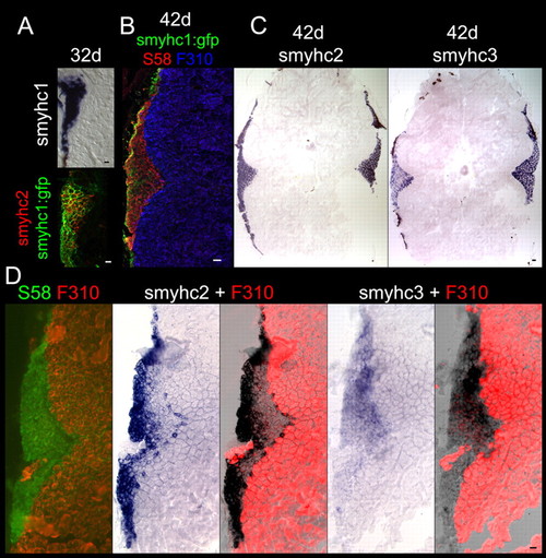
Expression of smyhc1, smyhc2 and smyhc3 in the juvenile and adult trunk. All panels show trunk transverse cryosections. (A) At 32 dpf smyhc1 (1 to 249 bp) in situ hybridisation shows expression in the lateralis superficialis, while smyhc2 (1 to 324 bp) in situ hybridisation shows colocalisation with smyhc1:gfp in the lateralis superficialis. (B) At 42 dpf, smyhc1:gfp shows colocalisation with slow MyHC S58 antigen in the lateralis superficialis, which is not fast muscle F310 antigen positive. (C) At 42 dpf, smyhc2 (1 to 324 bp) and smyhc3 (1 to 129 bp) in situ hybridisation shows expression in the lateralis superficialis. smyhc1 (1 to 249 bp) in situ hybridisation on adjacent sections failed to detect any expression. (D) Adjacent sections of 22 month post-fertilisation adult with smyhc2 (1 to 324 bp) and smyhc3 (1 to 129 bp) in situ hybridisation showing expression in subsets of the slow MyHC S58 antigen positive, fast muscle F310 antigen negative, lateralis superficialis. smyhc1 (1 to 249 bp) in situ hybridisation on adjacent sections failed to detect any expression throughout the trunk. Scale bars: 25 μm. EXPRESSION / LABELING:
|

Differential expression of Smyhc genes in craniofacial muscles. (A) Lateral and ventral views of the dorsal group of craniofacial muscles at 96 hpf with slow troponin C (stnnC), smyhc1 (1 to 249 bp), smyhc2 (1 to 324 bp) or smyhc3 (1 to 129 bp) in situ hybridisation (see Fig. 1A,D,E,G for gross location of these muscles). The lap and do lack smyhc1 expression while smyhc3 expression is restricted to the lap. (B) Ventral views showing ventral craniofacial muscles at 96 hpf with slow troponin C (stnnC), smyhc1 (1 to 249 bp), smyhc2 (1 to 324 bp) or smyhc3 (1 to 129 bp) in situ hybridisation. The ima, sh, iob and posterior tv lack smyhc1 expression. (C) Lateral and ventral views of the dorsal group of craniofacial muscles at 96 hpf showing colocalisation of slow MyHC S58 antigen with smyhc2 (1 to 324 bp) in situ hybridisation and, in the ao and lo, with smyhc1:gfp. (D) Ventral view at 96 hpf showing colocalisation of smyhc1:gfp and smyhc2 (1 to 324 bp) in situ hybridisation with slow MyHC S58 antigen. In the am, some fibres express smyhc2 and S58 antigen but not smyhc1:gfp (blue arrows). Some fibres express S58 antigen but neither smyhc2 nor smyhc1:gfp (white arrows). The extraocular muscles have zones of S58 antigen-expressing fibres that co-express smyhc1:gfp and zones that do not (pink arrows). (E) Ventral view at 96 hpf showing expression of slow troponin C in situ hybridisation (stnnC) in all slow MyHC S58 antigen-expressing fibres. Scale bars: 25 μm. Abbreviations: abh, abductor hyoideus; am, adductor mandibulae; ao, adductor operculi; do, dilator operculi; hh, hyohyoidus; ih, interhyoideus; ima, intermandibularis anterior; imp, intermandibularis posterior; io, inferior oblique; iob, inferior obliquus; ir, inferior rectus, lap, levator arcus palatini; lo, levatator operculi; sh, sternohyoideus; tv, transversus ventralis. EXPRESSION / LABELING:
|
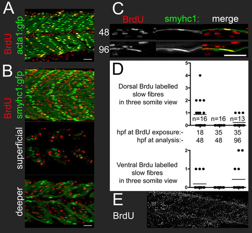
BrdU labelling shows that wild type embryos have smyhc1-expressing secondary superficial slow fibres. All panels show lateral views of posterior trunk, level with the anus. (A) At 48 hpf, embryos exposed to BrdU at 18 hpf show extensive BrdU co-labelling with acta1:gfp in nuclei that have the characteristic fast muscle elongated and diagonal morphology. (B) At 48 hpf, embryos exposed to BrdU at 18 hpf have BrdU-labelled nuclei superficial to the smyhc1:gfp-labelled surface slow fibres in the presumptive external cell layer (nuclei superficial to the surface slow fibres; these nuclei have a characteristic large flattened disk morphology) and immediately medial to the surface slow fibres, but not in the main body of the surface slow fibre layer (see also Movie 1). (C) BrdU and smyhc1:gfp co-labelling at dorsal somite margin at 48 hpf after BrdU exposure at 18 hpf, and at ventral somite margin 96 hpf after BrdU exposure at 35 hpf. (D) Scatter plots showing number of BrdU and smyhc1:gfp co-labelled nuclei in the dorsal or ventral myotome within a three-somite view at the level of the anus. Each dot represents the count in one embryo. The lines show the mean. Data are shown for exposure to BrdU at 18 hpf or 35 hpf, and with analysis at 48 hpf or 96 hpf. The BrdU labelled smyhc:gfp nuclei were always the smyhc1:gfp nuclei closest to the dorsal or ventral margin of the myotome. (E) At 48 hpf, embryos exposed to BrdU at 35 hpf have extensive labelling in cell types other than the surface slow fibres. Scale bars: 25 μm. EXPRESSION / LABELING:
|

Hedgehog dependent and independent smyhc1 and smyhc2 expression. All panels show lateral views. (A) smyhc1 (1 to 249 bp) in situ hybridisation at 48 hpf showing expression in secondary superficial slow fibres at the dorsal and ventral edges of the somites of a smo mutant. (B) smyhc2 (1 to 155 bp) in situ hybridisation at 48 hpf showing wild-type levels of expression in the iob of smo mutant (see Fig. S1 in the supplementary material for comparison with wild type). (C) Untreated and cyclopamine treated 55 hpf embryos showing Hh-independent smyhc1:gfp in the secondary superficial slow fibres and Hh-independent smyhc2 (1 to 324 bp) in situ hybridisation signal in the sca and iob. (D) 96 hpf wild-type sib and smo mutant. The smo mutant has slow MyHC S58 antigen in iob, sca and secondary superficial slow fibres; smyhc2 (1 to 324 bp) in situ hybridisation signal in iob and sca; but lacks scp and icp smyhc2 (1 to 324 bp) in situ hybridisation signal. (E) Untreated embryo and embryo exposed to cyclopamine from the 20-somite stage. This late cyclopamine exposure does not eliminate the smyhc1:gfp surface slow fibres but does eliminate smyhc2 (1 to 324 bp) in situ hybridisation signal in the scp and icp, as well as causing a dorsoventral narrowing of the posterior tail. Fifteen out of 15 embryos with this treatment showed this phenotype. (F) High-magnification views of posterior tail in untreated embryo and embryo exposed to cyclopamine from the 20-somite stage. In untreated embryos, smyhc2 (1 to 324 bp) in situ hybridisation signal colocalises with the general muscle MF20 antigen in the scp and icp dorsal and ventral to the smyhc:gfp surface slow fibre layer. The treated embryos lack scp and icp smyhc2 (1 to 324 bp) in situ hybridisation signal and general muscle MF20 antigen dorsal or ventral to the smyhc:gfp surface slow fibre layer. Scale bars: 25 μm. Abbreviations: icp, infracarinalis posterior; iob, inferior obliquus; sca, supracarinalis anterior; scp, supracarinalis posterior. |
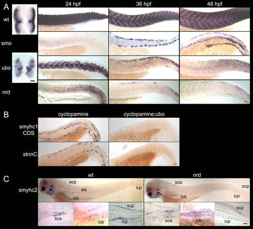
Hedgehog-independent secondary superficial slow fibres require Prdm1, whereas smyhc2 expression does not. (A) Lateral views of smyhc1-CDS in situ hybridisation on 24 hpf, 36 hpf and 48 hpf smo mutants, ubo (prdm1)tp39 mutants, nrd (prdm1)m805 and wild-type (wt) siblings; and transverse sections of 24 hpf ubo (prdm1)tp39 mutants and wild-type siblings. The near absence of Smyhc-expressing primary slow fibres in smo mutants at 24 hpf allows secondary slow fibres to be identified unambiguously at later stages. At 24 hpf, ubo (prdm1)tp39 or nrd (prdm1)m805 mutants have abundant, misplaced, weakly Smyhc-expressing fibres. Thus, it is unclear whether Smyhc-expressing fibres at later stages in prdm1 mutants are simply the remnants of those present at 24 hpf. (B) When primary slow fibre specification is prevented by cyclopamine treatment, 48 hpf genotyped ubo (prdm1)tp39 mutants lack almost all smyhc1-CDS in situ hybridisation and slow troponin C (stnnC) in situ hybridisation in secondary superficial slow fibres, whereas genotyped wild-type siblings have robust expression. (C) smyhc2 (1 to 155 bp) in situ hybridisation showing 96 hpf nrd (prdm1)m805 mutants have near wild-type levels of expression in the sca, iob, scp, icp and craniofacial muscles, but lack smyhc2-expressing els fibres. High magnification dorsal views show sca and lateral views show iob and scp/icp. Scale bars: 25 μm. Abbreviations: els, embryonic lateralis superficialis; icp, infracarinalis posterior; iob, inferior obliquus; sca, supracarinalis anterior; scp, supracarinalis posterior. |

The prdm1:gfp transgenic reporter marks primary and secondary slow fibres. (A) Weak prdm1 in situ hybridisation in presumptive secondary superficial slow fibre cells (arrows) at 35 hpf in smo mutants. (B) prdm1:gfp fluorescence in adaxial cells at the six-somite stage is almost eliminated in smo mutants. (C) At 24 hpf, Prox1-expressing primary slow fibres are marked with prdm1:gfp and are absent in smo mutants. (D) Lateral views of posterior trunk of 48 hpf smo mutants showing prdm1:gfp colocalised with smyhc1-CDS in situ hybridisation and slow troponin C (stnnC) in situ hybridisation in secondary superficial slow fibres at the dorsal and ventral edges of the somites. (E) In smo mutants, prdm1:gfp colocalises with Prox1 antigen in secondary fibres (arrows) in somite 13 at 36 hpf. Note the intense Prox1 staining in isolated cells that are external to the myotome (arrowheads). (F) At 48 hpf, in smo mutant posterior trunk, slow MyHC S58 antigen and prdm1:gfp mark secondary superficial slow fibres that do not colocalise with fast muscle F310 antigen. (G) Dorsolateral view of the iob and dorsal view of the sca at 96 hpf showing colocalisation of prdm1:gfp with slow MyHC S58 antigen but not with fast muscle F310 antigen (see Fig. 1 for gross location of these muscles). Scale bars: 25 μm. Abbreviations: iob, inferior obliquus; sca, supracarinalis anterior. EXPRESSION / LABELING:
PHENOTYPE:
|
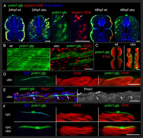
The prdm1:gfp transgenic reporter provides a lineage tracer to follow the fate of prdm1-dependent muscle precursors in ubo (prdm1) mutants. (A) Transverse sections show colocalisation prdm1:gfp with smyhc1-CDS in situ hybridisation in ubo (prdm1)tp39 mutants and wild-type siblings at 24 hpf. At 48 hpf, the prdm1:gfp marked muscle fibres are still present in ubo (prdm1)tp39 mutants, although Smyhc expression is reduced. Cell boundaries are marked with anti-β-catenin 15B8. (B) Lateral views showing that muscle fibres with prdm1:gfp form a superficial layer of horizontal, mononucleate fibres in 48 hpf wild-type embryos but are among the fast fibres in ubo (prdm1)tp39 mutants and like fast fibres are frequently multinucleate (TOTO stain) and orientated diagonally. (C) Transverse sections at 48 hpf show colocalisation of F310 fast muscle antigen with prdm1:gfp in ubo (prdm1)tp39 mutants but not in wild-type siblings. (D) In posterior trunk at 48 hpf, in ubo (prdm1)tp39 mutants, the dorsal fibres with persistent slow MyHC S58 antigen are marked with prdm1:gfp and fast muscle F310 antigen. (E) In posterior trunk at 36 hpf, in ubo (prdm1)tp39 mutants, even the most dorsally positioned fibres marked with prdm1:gfp lack Prox1 (arrows). Note the intense Prox1 staining in isolated cells external to the myotome (arrowheads). (F) In posterior trunk of cyclopamine-treated embryos, prdm1:gfp marked secondary fibres colocalise with slow MyHC S58 antigen but not fast muscle F310 antigen in genotyped wild-type siblings and vice versa in genotyped ubo (prdm1)tp39 mutants. Scale bars: 25 μm. |
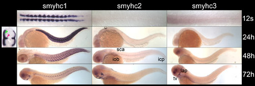
Onset of Smyhc gene expression in different muscles. ISH with smyhc1 (1 to 249 bp), smyhc2 (1 to 155 bp) or smyhc3 (1 to 129 bp) probes. Flat mounts of 12-somite stage embryos show adaxial cells express smyhc1 but not smyhc2 or smyhc3. Lateral views at 24 hpf, 48 hpf and 72 hpf show that smyhc2 is expressed from 48 hpf in the sca, iob and icp/scp, and smyhc3 is expressed in certain craniofacial muscles from 72 hpf. Transverse section of 24 hpf posterior trunk with smyhc1 (1 to 249 bp). ISH shows expression in the superficial slow fibres (green arrow) and muscle pioneers (pink arrow). Abbreviations: icp, infracarinalis posterior; iob, inferior obliquus; lap, levator arcus palatini; sca, supracarinalis anterior; scp, supracarinalis posterior; tv, transversus ventralis. EXPRESSION / LABELING:
|

slow troponin C is expressed in all S58 positive slow fibres and in the pectoral fin muscle. (A) At 96 hpf slow troponin C (stnnC) is expressed in the slow fibres throughout the embryo and also in pectoral fin muscle. (B) At 96 hpf, slow troponin C ISH shows co-localisation with slow MyHC S58 antigen in the smyhc1:gfp-positive superficial slow fibres and in the sca muscle (dorsal view anterior trunk), in els and iob muscles (lateral view anterior trunk), and in scp and icp muscles (lateral view posterior tail). The pectoral fin muscle expresses slow troponin C but not slow MyHC S58 antigen. Scale bars: 25 μm. Abbreviations: els, embryonic lateralis superficialis; icp, infracarinalis posterior; iob, inferior obliquus; pf, pectoral fin; sca, supracarinalis anterior; scp, supracarinalis posterior. EXPRESSION / LABELING:
|

smyhc1 expression in the craniofacial muscles is prdm1 independent. (A) Lateral views of wild-type sib and ubo (prdm1)tp39 mutant head at 96 hpf with immunofluorescence to the general muscle MF20 antigen and cartilage Col2 II-II6B3 antigen. In the ubo (prdm1)tp39 mutant, perturbations to the craniofacial muscles are associated with the major loss of skeletal elements in the posterior head mutant. The ceratobranchial cartilage is absent, and the sternohyoidus and inferior obliquus form abnormal attachments to the posterior basicranial commissure cartilage instead of the cleithrum. (B) Ventrolateral views of wild type and nrd (prdm1)m805 mutants at 96 hpf with smyhc1 (1 to 249 bp), smyhc2 (1 to 155 bp) or smyhc3 (1 to 129 bp) ISH showing prdm1-independent expression. nrd (prdm1)m805 mutants lack posterior transversus ventralis muscles. Scale bars: 25 µm. Abbreviations: bc, basicranial commissure; cb, ceratobranchial; iob, inferior obliquus; sh, sternohyoidus; tv, transversus ventralis. |