
- Title
-
Zebrafish blowout provides genetic evidence for Patched1-mediated negative regulation of Hedgehog signaling within the proximal optic vesicle of the vertebrate eye
- Authors
- Lee, J., Willer, J.R., Willer, G.B., Smith, K., Gregg, R.G., and Gross, J.M.
- Source
- Full text @ Dev. Biol.
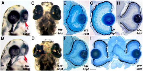
blowout mutants present with colobomas. Wild-type (A, C) and blw (B, D) mutant embryos at 50 hpf (A, B) and 5 dpf (C, D) imaged laterally (A, B) and ventrally (C, D). Colobomas can be unilateral or bilateral, and these blw mutants show bilateral incidence. Arrows in panels A and B point to the choroid fissure. Retinal tissue expelled from each eye has been outlined to illustrate the extent of the coloboma in panels C and D. Transverse histological sectioning of wild-type (E, G) and blw (F, H, I) eyes. Colobomas are obvious at 3 dpf in blw (F); however, at later time points their severity varies ranging from mild (H) to severe (I). Lens and retinal development appear normal in blw mutants. Anterior is up in panels A–D. Dorsal is up in panels E–I. Scale bars are 100 μm. |
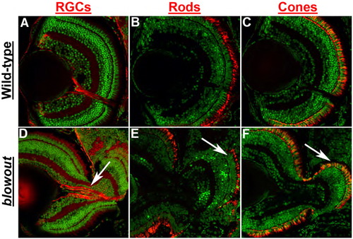
Retinal development is largely normal in blowout. Immunohistochemical analysis of retinal ganglion cells (RGCs) via zn8 staining (A, D), rod photoreceptors via zpr3 staining (B, E) and red/green double cones via zpr1 staining (C, F) in transverse retinal cryosections. All retinal cell types are present and are properly distributed in blw mutants, and retinal cell numbers are also similar between wild-type and blw mutant eyes. RGC organization is affected in blw mutants where RGC axons do not assemble into a tight bundle as they exit the eye (arrow in panel D). Photoreceptor outer segments are present in the retinal territories that are not contained within the eye cup (arrows in panels E, F). Antibody stains are red and nuclei are counterstained green with Sytox Green. Dorsal is up in all images. |
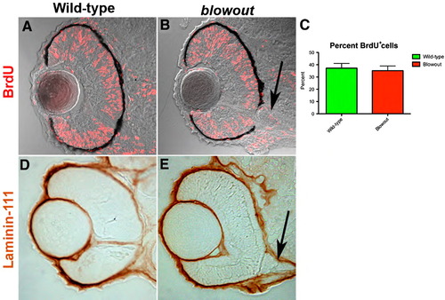
Retinal cell proliferation and Bruch’s membrane formation are both normal in blowout. (A, B) Wild-type (A) and blw (B) embryos were exposed to BrdU from 42 hpf to 48 hpf and immediately sacrificed. BrdU positive cells (red) are observed throughout the retinas of wild-type and blw mutant embryos in similar proportions (C; no statistical difference—Fisher’s exact test). Additional exposure periods (24–36 hpf, 72–96 hpf) resulted in identical regions of proliferation in the retinas of wild-type and blw mutants (data not shown). (D, E) Bruch’s membrane, a basement membrane at the posterior of the eye, highly expresses the laminin-111 protein (Lee et al., 2007). Shown here are images of 12 μm cryosections from wild-type (D) and blw (E) whole-mount embryos stained for laminin-111 protein at 48 hpf. Laminin-111 levels and distribution are similar between wild-type and blw embryos. The optic stalk appears abnormal in blw, as it remains connected to the retina at 48 hpf (arrows in panels B, E), while in wild-type siblings it has degenerated. Transverse sections, dorsal is up in all images. PHENOTYPE:
|
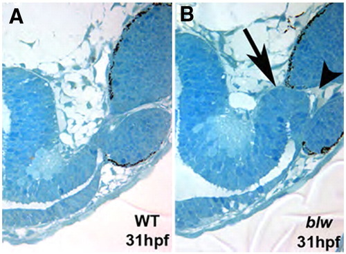
Optic stalk morphology is abnormal in blowout. Single 1.25 μm oblique histological sections taken from 31 hpf wild-type (A) and blw (B) embryos taken at the same plane of sectioning. The optic stalk in blw is larger and kinked at the distal end (arrow in panel B). Optic stalk tissue is present at the choroid fissure and appears to prevent the lateral edges of the fissure from meeting and fusing (arrowhead). PHENOTYPE:
|

The proximal/distal axis of the optic vesicle is disrupted in blowout. pax2a marks the proximal OV (optic stalk) in wild-type embryos at 15 hpf (A), 18 hpf (B), 24 hpf (G) and 36 hpf (H), while it is absent at 48 hpf (I) owing to the degeneration of the stalk by this time. Expression in blw mutants is observed in a broader domain within the optic stalk region as early as 15 hpf (B) and this expansion is maintained at 18 hpf (E), 24 hpf (J) and 36 hpf (K, arrow). Expression is not extinguished at 48 hpf and can be observed to extend into the retina (L, arrow). Conversely, pax6 marks the distal optic vesicle (retina and RPE) at 18 hpf (C), and expression is substantially contracted within distal optic vesicle of blw mutants (F) in a pattern complementary to the expanded domain of pax2a. EXPRESSION / LABELING:
PHENOTYPE:
|

Loss of ptc1 leads to colobomas and ventral optic cup defects. 1.3 ng injection of a translation blocking morpholino targeting ptc1 transcripts (ptc1MO) results in colobomas. Lateral views of a (A) Control morpholino (ControlMO) injected and (B) ptc1 morphant at 2.5 dpf. Morphants possess bilateral colobomas (arrow in panel B). Morphants are more severely affected than blw mutants, showing medial displacement and lateral flexure of the eyes, and lens displacement. Overt brain and muscle defects are also observed. (C–E) Transverse histological sections from a 5 dpf wild-type (C) and ptc1 morphants (D, E). Severe colobomas are obvious in the morphants (arrows in panels D, E), and in the many of them the lens is also displaced and lies out of the plane of the section. pax2a expression is expanded into the retina at 24 hpf (F, G) and pax6 expression is contracted (H, I). The Hh target genes ptc1 (J, K) and ptc2 (L, M) also show expanded domains of expression in ptc1 morphants. |

atp6v0b morpholinos do not phenocopy blowout. (A) The atp6v0b predicted genomic organization. atp6v0b MO targets the exon1/intron1 splice junction and leads to the inclusion of a 705 bp intron1 and the introduction of a premature stop codon. (B) RT-PCR validation of splice blocking efficacy of the atp6v0b MO. (C) Control injected and (D) atp6v0b morphant (13 ng) at 5 dpf. Morphants display oculocutaneous albinism and retinal degeneration but show no signs of colobomas even at the highest doses of MO injection. |
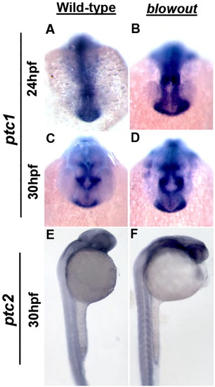
The domains of the Hh target genes ptc1 and ptc2 are expanded in blowout. Expression of ptc1 in the optic stalk of wild-type embryos at 24 hpf (A) and 30 hpf (C). (B, D) Expression in blw mutants is expanded and appears more intense at these time points both in the optic stalk, as well as in the brain and musculature (not shown). ptc2 expression is also expanded in blw mutants (F) relative to wild-type siblings (E). |

Adult homozygous blowout mutant phenotype. Two to three percent of homozygous blw mutants is viable and reveals additional roles for Patched1 in the formation of the adult zebrafish. (A) Lateral and (B) dorsal views of a heterozygous and (C, D) homozygous blw mutant fish at 1.25 years of age. blw mutants are smaller than wild-type embryos and show defects in craniofacial and/or jaw development where the lower jaw protrudes away from face. Defects are variable, but nearly all homozygous blw mutants display some degree of jaw malformations. (D) Homozygous mutants also display varying degrees of body curvature. PHENOTYPE:
|
Reprinted from Developmental Biology, 319(1), Lee, J., Willer, J.R., Willer, G.B., Smith, K., Gregg, R.G., and Gross, J.M., Zebrafish blowout provides genetic evidence for Patched1-mediated negative regulation of Hedgehog signaling within the proximal optic vesicle of the vertebrate eye, 10-22, Copyright (2008) with permission from Elsevier. Full text @ Dev. Biol.