FIGURE SUMMARY
Title

Identification of a novel retinoid by small molecule screening with zebrafish embryos

Authors
Sachidanandan, C., Yeh, J.R., Peterson, Q.P., and Peterson, R.T.
Source
Full text @ PLoS One

DTAB causes anterior-posterior axis defects.
a) Chemical structure of DTAB. b) Embryos treated with higher concentrations of DTAB showed more anterior-posterior patterning defects than those treated with lower concentrations. Zebrafish embryos were treated from 1hpf to 48hpf with different concentrations of DTAB. c) Embryos treated earlier in development, and thus for longer duration were more severely affected than those treated later. Embryos were exposed to 3 μM DTAB from different times of development to 48hpf and compared to embryos treated with the vehicle DMSO. Vehicle (DMSO) treated controls developed normally. All observations were made at 48hpf.

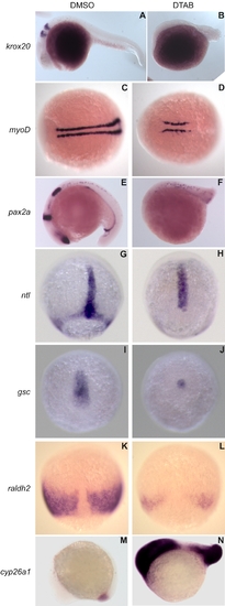
DTAB affects the expression of early developmental markers.
In situ hybridizations of zebrafish embryos show that treatment with DTAB results in severe reduction of krox20, pax2a, myoD, ntl, gsc, and raldh2 staining. DTAB treated embryos exhibited an upregulation of cyp26a1 transcripts in the tail and head. Embryos were treated with 12.5 μM DTAB or DMSO control from 4hpf onwards until fixation. Embryos were fixed at 75% epiboly (G–L), 10-somite (M–N), 18-somite (C–F) and 26-somite (A,B) stages. A–B, E–F, M–N are lateral views with anterior to the left and dorsal to the top. C–D are dorsal views with anterior to the left. G–L are dorsal views with the animal pole to the top.

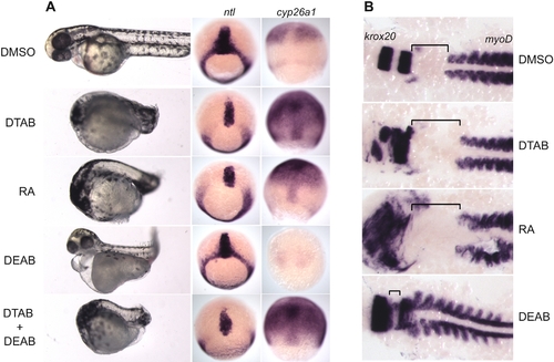
DTAB mimics effects of RA on early development.
a) Treatment with DTAB, RA and DTAB+DEAB results in anterior-posterior shortened phenotype. DEAB alone caused pericardial edema, twisted body axis and downregulation of cyp26a1. DEAB treatment did not affect the ntl staining while RA and DTAB treatment led to a loss of ntl staining in the dorsal margin. DTAB, RA and DTAB+DEAB caused an increase in cyp26a1 staining but DEAB alone caused downregulation of cyp26a1. Embryos were treated with DTAB (12.5 μM), RA (1 μM), DEAB (10 μM) and DTAB+DEAB or DMSO from 1hpf to 48hpf (for the bright field photography) and to 70% epiboly (for in situ hybridization for ntl and cyp26a1). Live bright field embryos are shown anterior to the left and dorsal to the top. In situ hybridized embryos, are shown, dorsal view with the animal pole to the top. b) DTAB and RA treatment caused an expansion in the distance between the krox20 and myoD positive domains. DEAB caused a compression of the ‘neck’ region and led to a decrease in the distance between krox20 and myoD domains. Embryos treated with DMSO, DTAB (1.6 μM), RA (0.125 μM) and DEAB (10 µM) were fixed at 9-somite stage and hybridized for krox20 (rhombomeres 3 & 5) and myoD (somites). Following in situ hybridization the embryos were de-yolked and flattened under a coverslip for better visualization.

Acknowledgments
This image is the copyrighted work of the attributed author or publisher, and ZFIN has permission only to display this image to its users. Additional permissions should be obtained from the applicable author or publisher of the image.
Open Access
Full text @ PLoS One